Showing posts with label Case Review Series. Show all posts

Obstetric & Gynecologic Ultrasound: Case Review Series 2nd edition

  • Section I: Opening Round.
  • Section II: Fair Game.
  • Section III: Challenge.


Obstetric & Gynecologic Ultrasound: Case Review Series 2nd edition by Karen L. Reuter and T. Kemi Babagbemi presents over 200 unknown cases-complete with over 350 state-of-the-art images, questions, answers, commentary, references, and more-to enhance your imaging interpretation skills in obstetric and gynecologic ultrasound. Discussions incorporate the most recent knowledge from OB/GYN ultrasound literature, providing an excellent review for both residents and practitioners.


Book features:
  • Follows the format of the Boards, and offers case studies similar to those likely to be found on exams, for a realistic preparation for the test-taking experience.
  • Presents cases in 3 overall categories-from least to most difficult-to build your skills in a cumulative way.
  • Offers cross references to Ultrasound: The Requisites, 2nd Edition so it's easy to find in-depth information on any subject.
  • Features over 350 state-of-the-art images-many of them new to this edition-that capture the full range of imaging findings in obstetric in gynecologic ultrasound.


New in this edition:
  • Places an increased emphasis on differential diagnosis, to help you distinguish specific diseases and disorders from others that have a similar sonographic presentation.
  • Includes new coverage of: 
    • abnormal IUD location,
    • chorioangioma of the placenta,
    • invasive molar pregnancy, 
    • fetal tachycardia causing non-immune fetal hydrops, 
    • vasa previa, 
    • fetal arachnoid cyst, 
    • amniotic sheet and amniotic band syndrome, 
    • postmenopausal ovarian cyst, 
    • autosomal recessive polycystic renal disease in the fetus, 
    • bladder flap hematoma, 
    • and more.
  • Groups cases by topic for a more efficient, targeted review of information.


Book Details

  • Paperback: 288 pages
  • Publisher: Mosby; 2 edition (October 13, 2006)
  • Language: English
  • ISBN-10: 0323039766 
  • ISBN-13: 9780323039765
  • Product Dimensions: 10.8 x 7.9 x 0.6 inches
List Price: $49.95 
 

Loevner: Brain Imaging: Case Review Series 2nd edition

  • 1 Acute Anterior Cerebral Artery Infarct.
  • 2 Cerebral Arteriovenous Malformation.
  • 3 Hemorrhagic Metastases - Melanoma.
  • 4 Penetrating Brain Injury – Bow and Arrow Injury.
  • 5 Orbital Pseudotumor.
  • 6 Acute Epidural Hematoma and Associated Linear Fracture of the Frontal Bone.
  • 7 Pyogenic Brain Abscess.
  • 8 Chiari II Malformation – with Associated Anomalies.
  • 9 Acute Hydrocephalus Secondary to Meningitis.
  • 10 Pineal Cyst.
  • 11 Hemorrhagic Contusions - Closed Head Injury.
  • 12 Juvenile Pilocytic Astrocytoma of the Cerebellum.
  • 13 Fibrous Dysplasia.
  • 14 Acute Middle Cerebral Artery Stroke: “Hyperdense” MCA and Insular Ribbon Sign.
  • 15 Multiple Sclerosis.
  • 16 Meningioma.
  • 17 Acute Actively bleeding Subdural hematoma – Subfalcine Herniation and Stroke.
  • 18 Acute Subarachnoid Hemorrhage - Rupture of an Anterior Communicating Artery Aneurysm.
  • 19 Embolic Infarcts (Acute and Subacute) – Atrial Fibrillation.
  • 20 Vestibular Schwannoma of the Internal Auditory Canal.
  • 21 Orbital Cellulitis and Abscess.
  • 22 Acute Hypertensive Thalamic Hemorrhage.
  • 23 Infiltrating Astrocytoma – Low Grade.
  • 24 Arachnoid Cyst .
  • 25 Retinoblastoma.
  • 26 Subdural Empyema – Complicated by Cerebritis.
  • 27 Glioblastoma Multiforme of the Corpus Callosum--"Butterfly Glioma"
  • 28 Virchow-Robin Perivascular Space
  • 29 Agenesis of the Corpus Callosum
  • 30 Nonaccidental Trauma--Child Abuse
  • 31 Cerebellar Hemangioblastoma
  • 32 Calvarial Metastases--Breast Carcinoma
  • 33 Chronic Anemia - Diffuse Replacement of the Fat in the Calvarial Marrow
  • 34 Giant Aneurysm – Middle Cerebral Artery
  • 35 Bilateral Subacute Subdural Hematomas
  • 36 Parafalcine Meningioma Invading the Superior Sagittal Sinus
  • 37 Hemorrhagic Venous Infarction
  • 38 Vertebral Artery Dissection - Spontaneous
  • 39 Spontaneous Cerebral Hematoma - Ruptured Cerebral Arteriovenous Malformation
  • 40 Vascular Infundibulum – Posterior Communicating Artery
  • 41 Closed Head Injury - Diffuse Axonal Injury
  • 42 Glioblastoma Multiforme – Subependymal Spread
  • 43 Amyloid Angiopathy
  • 44 Mesial Temporal Sclerosis
  • 45 Intraventricular Meningioma
  • 46 Suprasellar Germinoma
  • 47 Mineral Deposition in the Basal Ganglia on T1W Imaging – Abnormal Calcium/Phosphate Metabolism
  • 48 Herpes Simplex Encephalitis – Type 1
  • 49 Primary Orbital Lymphoma
  • 50 Basilar Meningitis and Encephalitis – Tuberculosis FAIR GAME
  • 51 Dural Metastases - Breast Carcinoma
  • 52 Aqueductal Stenosis
  • 53 Rathke's Cleft Cyst
  • 54 Optic Neuritis (Demyelinating Disease)
  • 55 Ocular trauma – Lens dislocation; Globe Rupture
  • 56 Developmental Venous Anomaly
  • 57 Fibrous Dysplasia of the Calvaria
  • 58 Cavernous Sinus Mass—Meningioma
  • 59 Pyogenic Ventriculitis With Acute Hydrocephalus
  • 60 Lesions of the Pituitary Stalk and Hypothalmus: Case A Sarcoidosis, Case B Langerhans Histiocytosis (EG)
  • 61 Nodular Subependymal Heterotopia
  • 62 Basilar Meningitis – Sarcoidosis
  • 63 Pituitary Microadenoma
  • 64 Multiple Sclerosis – Marburg Type
  • 65 Transtentorial Herniation
  • 66 Epidermoid Cyst - Cerebellopontine Cistern
  • 67 Cavernous Malformation (Cavernous Hemangioma, Occult Cerebrovascular Malformation)
  • 68 Persistent Trigeminal Artery
  • 69 Colloid Cyst
  • 70 Cavum Septum Pellucidum and Cavum Vergae
  • 71 Toxoplasmosis Infection in Acquired Immunodeficiency Syndrome
  • 72 Encephalocele - Developmental
  • 73 Cysticercosis of the Central Nervous System
  • 74 Facial nerve – Inflammation (Viral)
  • 75 MR Tractography – Preoperative Mapping of the Corticospinal Tracts for GBM Resection
  • 76 Right Posterior Inferior Cerebellar Artery Aneurysm Rupture
  • 77 Ganglioglioma
  • 78 Paget's Disease – Osteitis Deformans
  • 79 Fibromuscular Dysplasia – Complicated by Acute Stroke
  • 80 Carbon Monoxide Poisoning
  • 81 Lateral Medullary Syndrome – Wallenberg’s
  • 82 Wallerian Degeneration
  • 83 Dandy-Walker Malformation
  • 84 Thyroid Ophthalmopathy – with Optic Nerve Compression in Orbital Apex
  • 85 Bilateral Frontal Sinus Mucoceles
  • 86 Suprasellar Cistern Arachnoid Cyst
  • 87 Fourth Ventricular Neoplasms – Choroid Plexus Papilloma, and Ependymoma
  • 88 Multiple Cavernous Malformations - Familial Pattern
  • 89 Vein of Galen Aneurysm
  • 90 Tuberous Sclerosis (Bourneville's Disease)
  • 91 Craniosynostosis—Metopic Suture
  • 92 Glioma of the Tectum (Quadrigeminal Plate)
  • 93 Craniopharyngioma – Recurrent Adamantinomatous Type
  • 94 Intradiploic Epidermoid Cyst
  • 95 Porencephalic Cyst
  • 96 Subependymal Heterotopia
  • 97 Acute Middle Cerebral Artery Stroke - MR
  • 98 Hemangioblastoma
  • 99 Posterior Circulation Ischemia due to Subclavian Steal
  • 100 CNS Vasculitis – Cerebral Hemorrhage and Infarcts
  • 101 Optic Nerve Meningioma
  • 102 Peri-pineal Meningioma
  • 103 Air Embolism with Acute Right Cerebral Infarct
  • 104 Venous Sinus Thrombosis Complicating Otomastoiditis
  • 105 Intraventricular Hemorrage
  • 106 Pituitary Macroadenoma – in Left Cavernous Sinus
  • 107 Lymphoma of Occipital Condyle and C1
  • 108 Central Nervous System Sarcoidosis
  • 109 Normal-Pressure Hydrocephalus
  • 110 Lipomas Associated with the Corpus Callosum
  • 111 Sinonasal Undifferentiated Carcinoma with Intracranial Extension
  • 112 Intracerebral Rupture of a Posterior Communicating Artery Aneurysm
  • 113 Gadolinium-Associated Nephrogenic Systemic Fibrosis (Part I)
  • 114 Gadolinium-Associated Nephrogenic Systemic Fibrosis (Part II)
  • 115 Fenestration of the Basilar Artery
  • 116 Moyamoya Disease
  • 117 Focal Cortical Dysplasia
  • 118 Acute Sinusitis
  • 119 Anterior Clinoiditis and Mucocele Formation Complicating Sinusitis
  • 120 Spontaneous Intracranial Hypotension
  • 121 Multiple Meningiomas Following Remote Whole Brain Irradiation
  • 122 Fahr Disease (Familial Cerebrovascular Ferrocalcinosis)
  • 123 Central Nervous System Lymphoma--Immunocompetent Patient
  • 124 Alzheimer's Disease
  • 125 Sturge-Weber Syndrome (Encephalotrigeminal Angiomatosis)
  • 126 Ectopic Posterior Pituitary and Nonvisualization of the Infundibulum - Hypopituitarism
  • 127 Acute Toxic Demyelination – Carbon Monoxide Intoxication
  • 128 Neurofibromatosis Type 1 – Pilocytic Astrocytoma of Optic Pathway
  • 129 Lymphoma Involving the Circumventricular Organs
  • 130 Nonaneurysmal Perimesencephalic Subarachnoid Hemorrhage
  • 131 Radiation Necrosis Following Treatment of High-Grade Glioma
  • 132 Radiation Necrosis After XRT for Nasopharyngeal Cancer
  • 133 Internal Carotid Artery Dissection
  • 134 Tension Pneumocephalus
  • 135 Cryptococcosis
  • 136 Subarachnoid Seeding--Leptomeningeal Carcinomatosis
  • 137 Brainstem Astrocytoma
  • 138 Thrombosis of a Developmental Venous Malformation – Postpartum
  • 139 Textiloma or Gossypiboma - Retained Surgical Sponge
  • 140 Petrous Apex Cholesterol Granuloma
  • 141 Hamartoma of the Hypothalamus
  • 142 Hodgkin Disease – Dural Based
  • 143 Clival Chordoma
  • 144 Perineural Spread of Skin Cancer - Basal Cell Carcinoma
  • 145 Central Nervous System Lyme Disease
  • 146 Reversible Posterior Leukoencephalopathy (RPL) – PRES
  • 147 Fibrous Dysplasia of the Temporal Bone
  • 148 Neuroblastoma – Metastases to the Cranium
  • 149 Cystic Meningioma – with Cellular Atypia
  • 150 Global Anoxic Brain Injury
  • CHALLENGER
  • 151 Lobar Holoprosencephaly
  • 152 Olivopontocerebellar Degeneration
  • 153 Band Heterotopia—Pachygyria
  • 154 Huntington's Disease
  • 155 Meningocele/Pseudomeningocele of the Skull Base (Petrous Apex)
  • 156 Right Thalamic Glioblastoma Multiforme
  • 157 Multiple Sclerosis and Glioblastoma Multiforme
  • 158 Adrenoleukodystrophy
  • 159 Medulloblastoma
  • 160 Subacute Middle Cerebral Artery Territory Stroke Mimicking a Bone Metastasis
  • 161 Cytomegalovirus Meningitis and Ependymitis in a Patient with AIDs
  • 162 Upper Brainstem and Thalamic Cavernous Malformation
  • 163 Capillary Telangiectasia and Developmental Venous Anomaly of the Brain Stem
  • 164 Central Neurocytoma
  • 165 Marchiafava-Bignami Syndrome
  • 166 Metastatic Carcinoid to the Orbital Extraocular Muscles
  • 167 Joubert's Syndrome
  • 168 Systemic Non-hodgkin Lymphoma
  • 169 Abnormal Iron Deposition in Deep Gray Matter Nuclei
  • 170 Herpes Encephalopathy - HHV-6 Following Organ Transplantation
  • 171 Jacob-Creutzfeldt Disease
  • 172 Orbital Lymphangioma
  • 173 Carotid-Cavernous Fistula – Part I
  • 174 Carotid-Cavernous Fistula – Part II
  • 175 Gliomatosis Cerebri
  • 176 Canavan's Disease
  • 177 Neurofibromatosis Type 2
  • 178 Metastasis to the Cerebellopontine Angle - Ocular Melanoma
  • 179 Retained Gadolinium - Renal Failure
  • 180 Amyotrophic Lateral Sclerosis – Lou Gehrig’s Disease
  • 181 High Grade Anaplastic Astrocytoma
  • 182 Wilson's Disease
  • 183 Pleomorphic Xanthoastrocytoma
  • 184 Primary Leptomeningeal Melanosis
  • 185 Dural Arteriovenous Fistula (Part I)
  • 186 Dural Arteriovenous Fistula (Part II)
  • 187 Fetal Schizencephaly
  • 188 Wernicke Encephalopathy
  • 189 Ruptured Mycotic Aneurysm
  • 190 Osmotic Demyelination
  • 191 Rosai-Dorfman Disease – Benign Sinus Histiocytosis
  • 192 Cystic Necrotic Brain Mass – Part I
  • 193 Cystic Necrotic Brain Mass Part II– Metastatic Adenocarcinoma of the Lung
  • 194 Rhombencephalosynapsis
  • 195 Progressive Multifocal Leukoencephalopathy
  • 196 Sphenoid Dysplasia and Brain Dysplasia- Neurofibromatosis Type 1 (von Recklinghausen's Disease)
  • 197 Panhypopituitarism with Absence of the Infundibulum
  • 198 Progressive External Ophthalmoplegia Syndrome – Due to Mitochondrial Defect
  • 199 Limbic Encephalitis - Paraneoplastic Syndrome (Small Cell Lung Carcinoma)
  • 200 von Hippel-Lindau Disease


This volume in the best-selling "Case Review" series uses hundreds of case studies to challenge your knowledge of a full range of topics in brain imaging. With 170 brand new cases, new coverage of MRA, CTA, MR spectroscopy and multi-detectors and over 600 brilliant images, this is your ideal concise, economical, and user-friendly tool for self assessment in this specialty!


Book Features
  • Utilizes case studies organized into "Opening Round," "Fair Game," and "Challenge" sections, so you can test yourself at varying difficulty levels.
  • Provides at-a-glance review/self-testing of brain imaging cases ideal for preparing for the boards in brain imaging, the CAQ exam for neuroradiology or for the general radiologist ready for re-certification.
  • Mimics the official exam formats and daily practice environment by giving you cases/images as unknowns with three to four questions; then, on the flip side of the page, diagnosis, answers to the questions, additional commentary, and references to the corresponding volume in Elsevier's popular Requisites Series.
  • Includes 600 state of the art images to effectively compliment and support the text and provide a clear picture of what you can expect, both in test-taking and in practice.
  • Uses randomly organized cases so you can test yourself without the aid of logical organization by anatomy or disease type.


New in this edition
  • Includes 170 new cases and over 50 new diagnoses so you can keep pace with the latest developments.
  • Includes a greater emphasis on differential diagnosis.
  • Adds coverage of MRA, CTA, MR spectroscopy and multi-detectors to keep you completely current.
  • Provides all new images for existing entities.
  • Adds cutting-edge coverage of neuro-imaging including spectroscopy, CTA, MRA, Functional imaging, tractography, perfusion and diffusion.


Book Details

  • Paperback: 432 pages
  • Publisher: Mosby; 2 edition (November 13, 2008)
  • Language: English
  • ISBN-10: 032303179X
  • ISBN-13: 978-0323031790
  • Product Dimensions: 10.7 x 8.2 x 0.7 inches
List Price: $49.95 
 

Top 3 Differentials in Radiology: A Case Review

Top 3 Differentials in Radiology: A Case Review is a practical case-based reference that will enable radiologists and radiology residents to hone their skills in developing differential diagnoses for common imaging findings. Presented as unknowns, the cases are arranged into twelve main sections based on radiology subspecialties. The book presents each case as a two-page unit. The left page features clinical images and a brief description of the clinical presentation. The right page provides the key imaging finding, “Top 3” differential diagnoses, additional differential diagnoses, the final diagnosis, and imaging pearls. The final section of the book contains selected cases from all radiology subspecialties with distinctive imaging findings that should lead definitively to a single diagnosis.

Book Features:
  • 325 cases presented as unknowns to facilitate exam preparation
  • Valuable high-yield review of all disease entities on the list of differential diagnoses for each case
  • More than 700 high-quality images, including 74 in full color, depicting key radiographic findings
  • Imaging pearls at the end of each case that highlight key teaching points
With its emphasis on gaining a solid foundation in differential diagnoses for the full range of key imaging findings encountered in clinical practice, this book is ideal for individuals preparing for the initial American Board of Radiology examination as well as more experienced radiologists preparing for recertification examinations.

Product Details

  • Paperback: 720 pages
  • Publisher: Thieme; 1 edition (November 9, 2009)
  • Language: English
  • ISBN-10: 1604062266
  • ISBN-13: 978-1604062267
  • Product Dimensions: 11.1 x 8.8 x 1.2 inches 
List Price: $109.95

Schwartz Emergency Radiology: Case Studies

Emergency Radiology: Case Studies by David T. Schwartz is a one-of-a-kind text specifically designed to help you fine-tune your emergency radiographic interpretation and problem-solving skills. Illustrated with hundreds of high-resolution images, this reference covers the full range of clinical problems in which radiographic studies play a key role. Dr. David Schwartz, leading educator and expert author of Emergency Radiology, takes you step-by-step through the radiographic analysis of medical, surgical, and traumatic disorders, giving you an unparalleled review of the use and interpretation of radiographic studies in emergency diagnosis.

Book features:
  • 55 cases studies that highlight challenging areas in emergency diagnosis, including imaging studies with subtle, equivocal, or potentially misleading findings.
  • Detailed coverage of the broad spectrum of disorders for which radiographs are utilized in emergency practice.
  • Coverage of chest and abdominal radiology, the extremities, cervical spine and facial radiology, and head CT.
  • Cohesive template for each chapter, beginning with a case presentation, followed by a comprehensive discussion of the disorder under consideration.
  • Sections begin with an overview of the pertinent radiographic technique, anatomy, and method of radiographic interpretation.
  • Diagnosis-accelerating radiographs, ultrasound images, CT scans, and MR images.
  • Invaluable “pearls and pitfalls” of radiographic interpretation.
About the Author
David T. Schwartz, MD, FACEP, Associate Professor of Clinical Emergency Medicine, Bellevue Hospital and New York University Medical Center, New York, New York.

Book Details Emergency Radiology
  • Hardcover: 559 pages
  • Publisher: McGraw-Hill Professional; 1 edition (November 5, 2007)
  • Language: English
  • ISBN-10: 0071409173
  • ISBN-13: 9780071409179
  • Product Dimensions: 11 x 8.7 x 1.1 inches
  • List Price: $155.00
  • Price: $124.00
  • You Save: $31.00 (20%)
Buy Button

Genitourinary Imaging: Case Review Series 2nd edition

Genitourinary Imaging: Case Review Series 2nd edition by Ronald J. Zagoria, William W. Mayo-Smith and Julia R. Fielding provides an excellent case selection for sharpening diagnostic skills in this challenging subspecialty area. Emphasis is on differential diagnoses and pertinent radiological findings, but the relevant clinical points are also covered. This second edition contains numerous new images (more than 400 total images) as well as an addition of fifty cases. It is cross-referenced to GU RADIOLOGY: The Requisites, 2e.


Book features:
  • Volume in the Case Review series, covering brain imaging, targeted towards residents, fellows and practicing Radiologists.
  • Mimics official exam formats and daily practice environment with a pedagogically anchored format that introduces cases/images as unknowns with three to four questions; then, on the flip side of the page, diagnosis, answers to the questions, additional commentary, references and cross-references to The Requisites volume are supplied in logical succession; blank space is available for note taking.
  • Cases are randomly organized and then divided into three overall categories reflecting three different levels of difficulty.
  • Over 400 state of the art images.
  • Can be read cover to cover in a short period of time.


New in this edition:
  • All existing cases revised.
  • Approximately 50 new cases (including a greater focus on MR and Gyn).
  • Addition of a new co-editor (Fielding will supply the 50 new cases).
  • New images for approx. 75% of existing entities.
  • Greater emphasis on DDx.
  • Greater emphasis on patient treatment.
  • All references updated.


About the Authors
  • Ronald J. Zagoria, MD, Professor; Vice Chair for Medical Affairs; Section Head of Abdominal Imaging, Wake Forest University Baptist Medical Center, Winston-Salem, NC, US. 
  • William W. Mayo-Smith, MD, Associate Professor of Radiology, Brown University School of Medicine, Director of Computed Tomography, Rhode Island Hospital, Director of the Cross-Sectional Imaging Fellowship, Rhode Island Hospital / Brown University, Providence, RI. 
  • Julia R. Fielding, MD, Chief, Abdominal Imaging, Associate Professor of Radiology, University of North Carolina, Chapel Hill, NC, USA.


Book Details 
 
  • Paperback: 432 pages
  • Publisher: Mosby; 2 edition (September 25, 2006)
  • Language: English
  • ISBN-10: 0323037143
  • ISBN-13: 9780323037143
  • Product Dimensions: 10.8 x 8.3 x 0.8 inches
List Price: $49.95 
 

Breast Imaging: Case Review Series

Breast Imaging: Case Review Series by Emily Conant and Cecilia Brennecke reviews a full range of clinical issues in breast imaging, including mammographic positioning, calcifications, FNA, and ultrasound appearance of masses. Approximately 200 cases, with one to four unknown images and corresponding questions, probe your knowledge of a wide variety of topics in the field. Accompanying answers, commentary, and references help you to gain a better understanding of how the correct diagnosis was reached. Packed with hundreds of high-quality images, this affordable, easy-to-use resource offers an ideal way to sharpen your diagnostic skills and study for exams.


Book features:
  • Features a user-friendly case format to facilitate the quick location and review of specific information.
  • Divides cases into Opening Round, Fair Game, and Challenge categories allowing you to assess your proficiency at three different levels of complexity.
  • Includes a list of questions following each case - with answers and rationales - plus a brief discussion emphasizing characteristic imaging features, differential diagnostic considerations, and key points of clinical information.
  • Provides detailed commentary and selected helpful references on each topic.
  • Includes cross-references to Breast Imaging: The Requisites, by Debra Ikeda, MD.


About the Author
  • Emily Conant, MD, Associate Professor, Radiology Chief, Women’s Imaging University of Pennsylvania Medical Center, Philadelphia, PA.
  • Cecilia Brennecke, MD, Associate Professor, Radiology Johns Hopkins University, Director, American Radiology Services, Johns Hopkins at Green Spring, Lutherville, MD.


Book Details

  • Paperback: 232 pages
  • Publisher: Mosby; 1 edition (May 15, 2006)
  • Language: English
  • ISBN-10: 0323017460
  • ISBN-13: 9780323017466
  • Product Dimensions: 10.7 x 8.2 x 0.5 inches
List Price: $49.95 
 

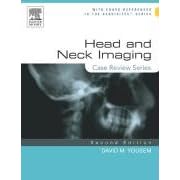
Yousem: Head and Neck Imaging: Case Review Series 2nd edition

Head and Neck Imaging: Case Review Series 2nd edition by David M. Yousem and Ana Carolina B. S. da Motta affords you a clinical tool that helps speed your differential diagnoses and ensures your proficiency. Organized like the Oral Boards, it serves as a study guide for exams, CAQ and re-certification-and as a personal review of the subspecialty. Here, Johns Hopkins' Drs. Yousem and Motta describe the latest techniques through 200 actual cases and 340 high-quality images. Tightly arranged, the text assures you fast access to information. Self-testing devices throughout validate your comprehension.

Book features:
  • Incorporates the most advanced imaging techniques.
  • Formatted like the Oral Board Exam for easy study and review.
  • Cross-referenced to Neuroradiology: The Requisites, Second Edition.
New in this edition:
    Obstetric and Gynecologic Ultrasound
  • Offers 200 all-new cases with new discussions and current literature references.
  • Presents over 340 images-including MR techniques (spectroscopy, diffusion, magnetization transfer, 3D FIESTA, tensor imaging) and CT techniques (CTA, bolus tracking, 3D reformats, perfusion imaging, Cine loops) and more.
  • Explores new topics including CT and MR angiography of the neck • multi-detector CT with 3D reconstructions • post-transplant lymphoproliferative disorders • new HIV infections • diagnostic and therapeutic image-guided procedures • medical economics • and much more.
  • Reflects a greater emphasis differential diagnosis and patient treatment.
Book Details 
  • Paperback: 336 pages
  • Publisher: Mosby; 2 edition (October 14, 2005)
  • Language: English
  • ISBN-10: 0323029892
  • ISBN-13: 9780323029896
  • Product Dimensions: 11 x 8.3 x 0.8 inches
List Price: $49.95

Copyright 2010 Radiology Book Review - All Rights Reserved.
Designed by Web2feel.com | Bloggerized by Lasantha - Premiumbloggertemplates.com | Affordable HTML Templates from Herotemplates.com.