Showing posts with label MRI. Show all posts

Berquist: MRI of the Musculoskeletal System 5th Edition

The Fourth Edition of MRI of the Musculoskeletal System text was published in 2001. Since that time, new magnet configurations (extremity, open) and higher field strengths (3 T) have begun to impact the practice for musculoskeletal magnetic resonance (MR) imaging. Coupled with improvements in software, pulse sequences, and examination techniques, the musculoskeletal applications for MR imaging have expanded dramatically. Gadolinium use for intravenous, intraarticular, and angiographic imaging has become routine. Spectroscopy in the clinical setting is also more commonly employed, though not to the level of more conventional imaging techniques.

The Fifth Edition of MRI of the Musculoskeletal System provides significant updates on principles, techniques, and applications for musculoskeletal imaging.
 
  • Chapter 1 reviews basic principles of physics, pulse sequences, and terminology, using an approach that is easy to read and comprehend. 
  • Chapter 2 provides essentials of interpretation with many new images and pulse sequences to explain the signal changes of pathologic tissues compared to signal intensity of normal tissue. 
  • Chapter 3 discusses safety issues, sedation, patient selection, patient positioning, coil selection, pulse sequences, and the uses of gadolinium for musculoskeletal imaging. 
  • Chapters 4, 5, 6, 7, 8, 9, 10 and 11 are anatomically oriented, with new anatomic MR images in each chapter. Improved images using new techniques the expanded applications are reviewed in each chapter. Pediatric applications are included in these anatomic chapters as appropriate.
  • A thorough discussion of musculoskeletal neoplasms and neoplasm-like conditions is included in Chapter 12. 
  • Chapter 13 is dedicated to musculoskeletal infections, including soft tissue, osseous, articular, spondylopathy, and postoperative infections. 
  • Chapter 14 provides in-depth coverage of diffuse marrow diseases. 
  • Chapter 15 is designed to review miscellaneous and evolving MR imaging applications. 
  • The final chapter, Chapter 16, updates clinical uses of spectroscopy.

New technology has led to a significant increase in the utility of MRI for evaluation of the musculoskeletal system. This edition provides a thorough review of anatomy, techniques, and applications, with many new images. This text will be of use to physicians, residents, students, and other health professionals who perform or request MR imaging examinations.

 
Book Details

  • Hardcover: 1008 pages
  • Publisher: Lippincott Williams & Wilkins; Fifth edition (October 3, 2005)
  • Language: English
  • ISBN-10: 0781755026
  • ISBN-13: 978-0781755023

List Price: $236.00 
 

Ellis: Human Sectional Anatomy: Atlas of Body Sections, CT and MRI Images 3rd edition

Successive authors of atlases on sectional anatomy have emphasized the value to the anatomist and the surgeon of being able to view the body in this dimension. It is always difficult to consider three dimensions in the mind’s eye; to be able to view the relationships of the viscera and fascial planes in transverse and vertical section helps to clarify the conventional appearances of the body’s structure as seen in the operating theatre, in the dissecting room and in the textbook.
The introduction of modern imaging techniques, especially ultrasound, computed tomography (CT) and magnetic resonance imaging (MRI), has enormously expanded the already considerable importance of sectional anatomy. The radiologist, neurologist, internist, chest physician and oncologist, as well as specialists in the various fields of surgery, have had to re-educate themselves in the appearances and relationships of anatomical structures in transverse and vertical section. Indeed, precise diagnosis, as well as the detailed planning of therapy (for example, the ablative surgery of extensive cancer) and of interventional radiology, often depends on the cross-sectional anatomical approach.
This atlas combines three presentations of crosssectional anatomy – that of the dissecting room, CT and MRI. The series are matched to each other as closely as possible on opposite pages. Students of
anatomy, surgeons, clinicians and radiologists should find the illustrations of anatomical cross-sections (obtained by the most modern techniques of preparation and photographic reproduction) and the equivalent cuts on imaging (obtained on state-of-the art apparatus) both interesting and rewarding.

Manning: Cardiovascular Magnetic Resonance 2nd edition

Cardiovascular magnetic resonance (CMR) is a medical imaging field that excites great interest because it combines superb image quality with new techniques for probing the cardiovascular system in novel ways. What surprises is the versatility of the technology: blood flow, angiography, assessment of atherosclerosis, myocardial perfusion, focal necrosis, oxygen saturation, technology, and chemical composition are among the measurements that are being refined for clinical use, in addition to the well-known “gold standard” capabilities of CMR in defining anatomy and ventricular function. Such potential comes at a price, however, as this technology is not quickly learned, and highquality clinical practice needs experience. Professional didactic and clinical training is required for all newcomers to the field and to maintain cutting-edge competency.
The aim of this book is to provide instruction in the current clinical practice of CMR, while also highlighting areas of clinical potential, which are presently in varying stages of development. If we succeed in drawing new investigators and clinicians to enter the field or illuminating new areas for those already involved, then we will have achieved our objective—the healthy growth of competent and motivated practitioners in CMR for the benefit of clinical science, patient care, and the advancement of the field.

Atlas of Fetal and Postnatal Brain MR

It became obvious in the late 1990s that magnetic resonance (MR) imaging of the fetal central nervous system was going to be more than an intellectual curiosity wrapped around a technical challenge. There was (and remains in some circles) some resistance to accept that there is any need for supplementing ultrasonography with fetal MR in cases of suspected developmental brain abnormalities. Many recent studies have shown value of in utero MR of the fetus and there is also gathering interest in postmortem MR of the fetus as an adjunct or replacement to autopsy. The problem was how to start. Few radiologists have experience of the normal MR appearances of the brain at 20 to 40 weeks gestational age. Those who do have the experience have usually gained it from imaging premature babies in whom the predominant pathologies are the complications of prematurity, not malformations. It has taken us a long time to build up a base of normal fetal brain examinations; therefore our appreciation of age-related normality was slow to form. This atlas will help others in this complex area of image interpretation.

MR Angiography of the Body Technique and Clinical Applications

Indirect angiography following intravenous bolus injection of iodinated contrast media entered clinical practice as an alternative less-invasive method to direct (arterial) angiography when digital substraction X-ray techniques became available.
Because of its technical shortcomings, IVDSA was later replaced by CT angiography as another less-invasive modality for vascular imaging. The introduction of multislice CT scanners and rapid progress in electronic image reconstruction allowed CT angiography to become the actually well-known, highly performing, and reliable radiological modality.
MR angiography offers better patient safety than CT angiography because of the absence of ionizing irradiation. As compared with ultrasound vascular imaging, it offers and superior accuracy as well as better contrast resolution.
This book offers a comprehensive overview of the latest technological advances in MR angiography, including up to date image acquisition techniques as well as optimal sequences. Special attention is given to fl ow-based angiography, optimal and safe use of MR contrast media, rapid and reliable image procession, as well as to the correct interpretation of common artifacts.
The main section of this book is devoted to the current clinical applications of MR angiography in the different body organs and anatomic regions as well as to the important role of this method in the managing of transplant patients. The book is exquisitely illustrated with multiple high-quality demonstrative cases and as always presented in technically impeccable printing and lay out The editors are internationally well known for their superior and longstanding knowledge in MR angiography and image procession. They are supported by a group of distinguished collaborating authors, who are also experts in the field. It is highly recommended to general and organ specialized radiologists as a most welcome update of their knowledge and as a practical guide in their daily practice. This book may help practicing radiologists in making the best use of this exciting methodology, and encourage referring physicians to include MRA in the diagnostic work-up of their patients.

Contents
Image Acquisition Technique and Sequences
1 Flow-Based MRA
2 MR Angiography Contrast Agents
3 Image Acquisition Technique and Sequences Contrast-Enhanced MRA
4 Artifacts in MR-Angiography
5 Image Processing
Clinical Applications
6 Radiologic Vascular Anatomy
7 Neck Vessels
8 Thoracic Aorta and Pulmonary Vessels
9 Heart and Coronary Arteries
10 Abdominal Aorta and Renal Arteries
11 Peripheral Vessels
Special Topics: Transplants
12 MRA in Liver and in Orthotopic Liver Transplants
13 MRA in Transplanted Pancreas and Kidneys
Subject Index
List of Contributors

Product Details

  • Hardcover: 178 pages
  • Publisher: Springer; 1 edition (December 3, 2009)
  • Language: English
  • ISBN-10: 3540797165
  • ISBN-13: 978-3540797166
  • Product Dimensions: 10.8 x 7.8 x 0.6 inches
List Price: $239.00

Prayer: Fetal MRI (Medical Radiology / Diagnostic Imaging)

Driven by the ongoing technical and scientific progress, fetal MR imaging has evolved from an exclusively experimental prenatal imaging modality to a clinically important tool, which impacts decision making in the field of pre- and perinatal medicine. Nowadays, specialists involved in prenatal diagnosis are challenged with the demand to perform fetal MR examinations without the availability of basic reference with respect to methods, indications, MR appearance of maturing fetal and extrafetal organs, and pathological changes of intrauterine contents.
This book is the first handbook to offer a state-of-the art and comprehensive overview of the meticulous examination techniques and the specific sequences, which are essential for optimal image quality in fetal imaging. It further covers in depth all its current clinical applications as well as the safety issues.
The aim of this book is to provide basic information about fetal MRI not only for the examiners but also for the referring clinicians. MR methods are described in detail and safety aspects are discussed. The prerequisites of fetal MRI are described including patient preparation as well as the appropriate selection of MR parameters for the respective clinical questions. In order to fully exploit the potential of this modality, the MR characteristics of normal and pathological organ development are described on various MR sequences, including advanced MR imaging techniques with illustration by numerous figures. The possibilities of intrauterine surgical therapy are described using the example of treatment of congenital diaphragmatic hernia. For better understanding of normal and pathological brain development, the histological basis of early human brain maturation is provided. Different aspects of fetal neuroimaging are discussed comprising ultrasound and MR approaches.
Information that may help with interpretation of MR findings and their possible prognostic significance are summarized in chapters on genetics, maternal disease with possible impact on pregnancy, and postmortem MRI. In addition advice is given on how to treat postmortem specimens in order to obtain useful diagnostic clues.
All authors have been working scientifically and practically in the field. Thus the reader should receive well-grounded information on each topic.

BOLD fMRI: A Guide to Functional Imaging for Neuroscientists

Functional magnetic resonance imaging (fMRI) represents one of the most advanced and enlightening functional imaging techniques that has ever been developed. One major area of research interest in fMRI is within the field of Cognitive Neuroscience, which focuses on understanding all aspects of the mental processes involved in awareness, reasoning, and acquisition of knowledge and behavior. This book includes selected chapters from Functional MRI: Basic Principles and Clinical Applications (S. Faro and F. Mohamed, Eds. New York: Springer Science+Business Media, LCC 2006) that the editors feel are of particular interest to neuroscientists, as the focus is primarily on describing the basic principles of Blood Oxygen Level Dependent (BOLD) imaging and the developing clinical applications of fMRI in the neurosciences.
The first section of the book is an introduction to the physics principles of BOLD imaging as well as a review of fMRI scanning methodologies, data analysis, experimental design, and clinical challenges. The second section reviews some current and future clinical applications of fMRI, including the clinical fields of Language, Memory, fMRI WADA, and Brain Mapping. The third and final section is a pictorial neuroanatomical atlas of the basic motor, sensory, and cognitive activation sites within the brain. This section will give neuroscientists a familiarity with some of the more clinically relevant brain activation sites that are discussed in other chapters.
There has been a discovery of a tremendous body of knowledge in the relatively young field of fMRI.

Magnetic Resonance Imaging in Orthopedic Sports Medicine

Magnetic Resonance Imaging in Orthopedic Sports Medicine  is a practical resource on orthopedic MR imaging that bridges the backgrounds of radiologists and orthopedic surgeons. Radiologists learn why surgeons order imaging studies. They also learn terminology that will help them tailor reports to the specialty. Orthopedic surgeons gain insight on when to order an MRI, how MRI affects decision making, and how to interpret images. Case studies also depict key clinical and exam points, supplemented by MR images and illustrations. Shorter sections highlight other anatomical areas, and additional chapters address diagnostic accuracy and imaging pitfalls.
Though magnetic resonance imaging has helped revolutionize the field of orthopedic medicine, a difference in perspective persists between radiology and orthopedic specialists. Magnetic Resonance Imaging in Orthopedic Sports Medicine is an interdisciplinary resource designed to bridge this gap. Radiologists will gain insight into why surgeons order particular imaging studies, and orthopedic surgeons will learn when to order an MRI and how to best interpret its results. An orthopedic expert and a radiology expert weigh in on every chapter to provide in-depth analysis of the spine, shoulder, elbow, wrist, hand, hip, knee, ankle and other topics. Each subject is further examined in case studies, algorithms, flow charts, anatomical illustrations and hundreds of sample MR images. Magnetic Resonance Imaging in Orthopedic Sports Medicine is the only reference that combines the specialized knowledge of radiologists and orthopedic sports medicine physicians to accomplish the goal of better patient care.

Jackson: Dynamic Contrast-Enhanced Magnetic Resonance Imaging in Oncology (Medical Radiology / Diagnostic Imaging)

  • An Overview of Contrast-Enhanced MRI in Oncology.
  • Contrast Agents for Magnetic Resonance Imaging.
  • The Role of Blood Pool Contrast Agents in the Study of Tumor Pathophysiology.
  • Dynamic Susceptibility Contrast-Enhanced MRI in Oncology.
  • Quantification of Contrast-Enhancement in Dynamic Studies.
  • Pharmacokinetic Modelling of Contrast Kinetics in Dynamic MR Studies.
IMAGING TECHNIQUES:
  • Imaging Techniques for Dynamic Contrast-Enhanced Imaging.
  • Consensus Recommendations for Imaging in Angiogenesis.
CLINICAL APPLICATIONS:
  • Dynamic Magnetic Resonance Imaging in Cerebral Malignancy.
  • Dynamic Magnetic Resonance Imaging in Breast Tumours.
  • Dynamic Contrast Imaging in Cervical Carcinoma.
  • Dynamic Contrast-Enhanced Imaging in the Prostate.
  • Contrast-Enhanced MR Imaging in Musculoskeletal Tumors.
  • Contrast-Enhanced MR Imaging in the Liver.
APPLICATIONS:
  • The Use of Dynamic Contrast-Enhanced MRI Multicentre Trials.
  • Applications of Dynamic Magnet Resonance in Drug Development.
Subject Index.
List of Contributors.


Dynamic contrast-enhanced MRI is now established as the methodology of choice for the assessment of tumor microcirculation in vivo. This is assisting clinical practitioners in the management of patients with solid tumors and is finding prominence in the assessment of tumor treatments, including anti-angiogenics, chemotherapy, and radiotherapy.

In this book, targeted at both clinical practitioners and basic scientists, the principles of the methods, their practical implementation, and their application to specific tumor types are discussed by the leading authorities in the field today. The book will serve as an invaluable single-volume reference covering all the latest developments in contrast-enhanced oncological MRI.


About the Author
  • Alan Jackson, Professor of Neuroradiology at the University of Manchester, UK.
  • David Buckley, Lecturer in Magnetic Resonance Imaging at the University of Manchester, UK. Previous positions at the Universities of Florida, USA and Hull, UK.
  • Geoff Parker, Lecturer in Imaging Science at the University of Manchester, UK. Previous positions at the Institute of Neurology, London, UK and the Institute of Cancer Research, London, UK.


Book Details 
 
  • Paperback: 311 pages
  • Publisher: Springer; 1st edition (January 27, 2011)
  • Language: English
  • ISBN-10: 3642076076
  • ISBN-13: 978-3642076077
  • Product Dimensions: 10.2 x 7.5 x 0.8 inches
List Price: $239.00 

Cardiovascular MRI in Congenital Heart Disease - An Imaging Atlas (Springer) 2010

The last 10 years has seen explosive expansion of the number of centres performing cardiovascular magnetic resonance (CMR) imaging. The majority of this expansion has been in the field of adult ischaemic imaging, but congenital heart disease remains one of the main indications for CMR. Importantly, the greatly improved survival of patients with congenital heart disease gives us a burgeoning adult population living with the sequelae of the disease (grown-up congenital heart disease – GUCH).
Without previous experience or formal training, the interpretation of CMR images of patients with congenital heart disease can be difficult. The main aim of this book is to create a portable resource that offers effi cient access to high-quality MR (and where appropriate, CT) images of the common congenital and structural heart abnormalities. By providing key images for each condition and a clear interpretation of the MR appearances, will improve the reader’s understanding of the conditions, facilitate their interpretation of images and optimise the planning of the imaging protocols during their own practice of congenital CMR.
As with any publication from a single institution, the contents of this book represent the author's practice. But, this book is not a definitive or exhaustive description of the conditions. It is a factual, simple and eye-pleasing guide for fellows training in CMR, radiographers and technicians performing CMR scans, physician users of CMR, and perhaps those few in adult ischaemic practice, who may need the occasional aide memoir for incidental findings! This book useful in your everyday practice and learning.

Contents
1 Technical Considerations
2 MR Imaging Under GA
3 Imaging Protocol
4 Normal Anatomy-Axial
5 Normal Anatomy-Coronal
6 Normal Anatomy-Sagittal
7 Image Planes-Ventricles
8 Imaging Planes-Left Ventricular Outfl ow Tract
9 Imaging Planes-
Right Ventricular Outfl ow Tract
10a Imaging Planes-Branch PAs
10b Imaging Planes-Thoracic Aorta
11a Imaging Planes-Tricuspid Value
11b Imaging Planes-Mitral Value
12 Imaging Planes-Coronary Arteries
13 Atrial Septal Defect
14 Sinus Venosus Defect
15 Atrioventricular Septal Defect
16 Ventricular Septal Defect
17 Aortic Valve Stenosis
18 Aortic Valve Incompetence
19 Coarctation of the Aorta
20 Repaired Coarctation of the Aorta: Complications
21 Interrupted Aortic Arch
22 Aortic Vascular Rings
23 Left Pulmonary Artery Sling
24 Marfan Syndrome
25 Williams Syndrome
26 Mitral Stenosis
27 Mitral Regurgitation
28 Hypertrophic Cardiomyopathy
29 Dilated Cardiomypathy
30 Noncompaction Cardiomyopathy
31 Tetralogy of Fallot
32 Tetralogy of Fallot: Repaired
33 Pulmonary Stenosis
34 Percutaneous Pulmonary Valve Implantation
35 Pulmonary Atresia and VSD
36 Transposition of the Great Arteries: Arterial Switch Operation
37 Transposition of the Great Arteries: Senning and Mustard Repair
38 TGA with VSD and PS
39 Congenitally Corrected Transposition of the Great Arteries
40 Common Arterial Trunk
41 Double Outlet Right Ventricle
42 Double Inlet Left Ventricle
43 Hypoplastic Left Heart Syndrome: Norwood Stage 1
44 Bi-directional Cavo-pulmonary (Glenn) shunt
45 Fontan-Type Circulation (Tricuspid Atresia)
46 Total Cavo-pulmonary Connection
47 Anomalous Coronary Arteries
48 Anomalous Left Coronary Artery from Pulmonary Artery
49 Kawasaki Disease
50 Total Anomalous Pulmonary Venous Drainage
51 Partial Anomalous Pulmonary Venous Drainage
52 Ebstein’s Anomaly
53 Right Isomerism
54 Left Isomerism
55 List of Abbreviations
56 Further reading

Koh: Diffusion-Weighted MR Imaging - Applications in the Body

Radiological practice often evolves in small incremental steps rather than by quantum leaps. Old techniques or ideas may be given a new lease of life through novel applications, and anecdotes of these can be drawn from oncological imaging. For example, although positron emitting radionuclear tracers have been in use since the 1950s, it is only in the last decade that the power of positron emission tomography (PET) has been widely harnessed, such that PET-CT imaging is now mainstream and indispensible for the management of a patient with cancer. By comparison, although diffusion-weighted MR imaging (DW-MRI) has been routinely employed for the evaluation of intracranial diseases for two decades, it is only in the last few years that MR technological advances have enabled the technique to be successfully implemented in the body.
DW-MRI is appealing as an imaging technique for several reasons. First, the imaging can be performed relatively quickly and thus has the potential to be widely generalized and adopted. Second, the technique does not require the administration of exogenous contrast medium, which is attractive in the light of potential serious adverse effects of gadolinium-based contrast media. Third, the technique yields both qualitative and quantitative information, the latter being of particular importance as development of quantitative imaging techniques is now acknowledged to be critical to the future of radiology.
DW-MRI yields unique information that refl ects microstructural and functional alterations in tissues. Although there are still many challenges ahead, early experience with the technique has shown substantial promise. We hope that this volume will demonstrate the exciting potentials of this technique, discussing the applications of DW-MRI along broad themes of clinical practice rather than by organ systems.

Cardiovascular MRI in Congenital Heart Disease - An Imaging Atlas (Springer) 2010

The last 10 years has seen explosive expansion of the number of centres performing cardiovascular magnetic resonance (CMR) imaging. The majority of this expansion has been in the field of adult ischaemic imaging, but congenital heart disease remains one of the main indications for CMR. Importantly, the greatly improved survival of patients with congenital heart disease gives us a burgeoning adult population living with the sequelae of the disease (grown-up congenital heart disease – GUCH).
Without previous experience or formal training, the interpretation of CMR images of patients with congenital heart disease can be diffi cult. The main aim of Cardiovascular MRI in Congenital Heart Disease - An Imaging Atlas is to create a portable resource that offers efficient access to high-quality MR (and where appropriate, CT) images of the common congenital and structural heart abnormalities. By providing key images for each condition and a clear interpretation of the MR appearances, it will improve the reader’s understanding of the conditions, facilitate their interpretation of images and optimise the planning of the imaging protocols during their own practice of congenital CMR.
As with any publication from a single institution, the contents of this book represent the authors' practice. But it's not a definitive or exhaustive description of the conditions. However, This book is a factual, simple and eye-pleasing guide for fellows training in CMR, radiographers and technicians performing CMR scans, physician users of CMR, and perhaps those few in adult ischaemic practice, who may need the occasional aide memoir for incidental findings!

Contents
1 Technical Considerations
2 MR Imaging Under GA
3 Imaging Protocol
4 Normal Anatomy-Axial
5 Normal Anatomy-Coronal
6 Normal Anatomy-Sagittal
7 Image Planes-Ventricles
8 Imaging Planes-Left Ventricular Outflow Tract
9 Imaging Planes-Right Ventricular Outflow Tract
10a Imaging Planes-Branch PAs
10b Imaging Planes-Thoracic Aorta
11a Imaging Planes-Tricuspid Value
11b Imaging Planes-Mitral Value
12 Imaging Planes-Coronary Arteries
13 Atrial Septal Defect
14 Sinus Venosus Defect
15 Atrioventricular Septal Defect
16 Ventricular Septal Defect
17 Aortic Valve Stenosis
18 Aortic Valve Incompetence
19 Coarctation of the Aorta
20 Repaired Coarctation of the Aorta: Complications
21 Interrupted Aortic Arch
22 Aortic Vascular Rings
23 Left Pulmonary Artery Sling
24 Marfan Syndrome
25 Williams Syndrome
26 Mitral Stenosis
27 Mitral Regurgitation
28 Hypertrophic Cardiomyopathy
29 Dilated Cardiomypathy
30 Noncompaction Cardiomyopathy
31 Tetralogy of Fallot
33 Pulmonary Stenosis
34 Percutaneous Pulmonary Valve Implantation
35 Pulmonary Atresia and VSD
36 Transposition of the Great Arteries: Arterial Switch Operation
37 Transposition of the Great Arteries: Senning and Mustard Repair
38 TGA with VSD and PS
39 Congenitally Corrected Transposition of the Great Arteries
40 Common Arterial Trunk
41 Double Outlet Right Ventricle
42 Double Inlet Left Ventricle
43 Hypoplastic Left Heart Syndrome: Norwood Stage 1
44 Bi-directional Cavo-pulmonary (Glenn) shunt
45 Fontan-Type Circulation (Tricuspid Atresia)
46 Total Cavo-pulmonary Connection
47 Anomalous Coronary Arteries
48 Anomalous Left Coronary Artery from Pulmonary Artery
49 Kawasaki Disease
50 Total Anomalous Pulmonary Venous Drainage
51 Partial Anomalous Pulmonary Venous Drainage
52 Ebstein’s Anomaly
53 Right Isomerism
54 Left Isomerism
55 List of Abbreviations
56 Further reading

Kauczor: MRI of the Lung (Medical Radiology / Diagnostic Imaging Series)

One of the aims of the book series “Medical Radiology – Diagnostic Imaging” is to publish novel work that explores new frontiers in the field of radiological imaging. The investigation of the potential application of MRI to the study of lung diseases is a challenging and exciting field of clinical research driven by the rapid development of new techniques and sequences for cardiac MRI as well as by the use of noble hyperpolarised gases as novel contrast agents. The specific ability of MRI to visualize simultaneously morphological changes and modifications in perfusion, ventilation and gas exchange has opened up totally new perspectives in lung imaging. MRI of the lung thus eminently illustrates the mutation of the discipline of radiology from pure morphology to combined morphological-functional imaging.
The editor, H-U. Kauczor, is known internationally for his original research in various fields, and particularly into new applications of CT and MRI in oncology and lung pathology. For many years he has been the inspiration and principal investigator of a leading and innovative radiological research group in Heidelberg. Their original ideas and dynamic work organisation have resulted in an impressive list of scientific publications on various applications of MR imaging of the lung.

Contents
Technical

  • 1 General Requirements of MRI of the Lung and Suggested Standard Protocol
  • 2 MR Angiography of the Pulmonary Vasculature
  • 3 MR Perfusion in the Lung
  • 4 MRI of Pulmonary Ventilation
  • 4.1 Hyperpolarised 3He MRI: Physical Methods for Imaging Human Lung Function
  • 4.2 Hyperpolarized 129Xenon MRI of the Lung
  • 4.3 Fluorine MRI
  • 4.4 Proton MRI: Oxygen-enhanced Lung MRI and Alternative Approaches
  • 5 Respiratory Mechanics and Pulmonary Motion
Clinical
  • 6 Pulmonary Hypertension and Thromboembolic Disease
  • 7 Vascular Anomalies and Diseases
  • 8 Asthma
  • 9 Chronic Obstructive Pulmonary Diseases
  • 10 Cystic Fibrosis
  • 11 Lung Cancer
  • 12 Mediastinal Disease
  • 13 Pulmonary Infections – Pneumonia
  • 14 Interstitial Lung Disease
  • 15 Diseases of the Pleura and the Chest Wall 

Product Details
 
Paperback: 318 pages

Publisher: Springer; 1st edition (November 30, 2010) 

Language: English

ISBN-10: 364207099X / ISBN-13: 978-3642070990

List Price: $109.00 

CT and MR Angiography of the Peripheral Circulation - Practical Approach with Clinical Protocols by Debabrata Mukherjee (Informa Healthcare) 2007

The advent of multislice computed tomographic angiography (CTA) and magnetic resonance angiography (MRA) has had a dramatic impact on practice patterns in cardiovascular medicine with a significant proportion of catheterbased diagnostic angiography giving way to these new modalities.
This text represents a comprehensive treatise on CTA and MRA for the diagnosis of peripheral vascular disease and represents a consolidated attempt at bridging the everwidening gap between innovation in these areas and the knowledge of the general and cardiovascular clinician. A large number of high-quality illustrations make this textbook particularly attractive to the general clinician. Abundant tables and scanning protocols are included in each chapter wherever applicable.
In the first half of the book dedicated to CTA, the introductory chapters address technical principles, image reconstruction, radiation dosimetry, and principles of CT contrast agents and their administration. Subsequently, clinical application of CTA in each major vascular territory is dealt with thoroughly, with clear description of the examination and scanning technique for assessing each specific territory. The section on MRA provides a state of the art overview of this methodology in the peripheral circulation. MR physics relevant to the clinician is succinctly articulated with specific chapters dedicated to pulse sequences commonly used in peripheral angiography and MR contrast agents. Individual chapters on each major vascular territory including the extracranial and intracranial circulation, the pulmonary circulation, the thoracic and the abdominal aorta, the renal, and mesenteric circulation, the lower and upper extremity circulation enable the reader to appreciate the intricacies of MRA techniques for the evaluation of these territories.
In summary, Doctors Debabrata Mukherjee, Sanjay Rajagopalan, Informa Healthcare and colleagues have put together a very didactic and practical book. I believe the information will serve to stimulate the interest not only of the cardiovascular specialist ready to become familiarized with this important technological development of CT and MRI as applied to patients with peripheral vascular disease, but also of a broad range of professionals and students of cardiovascular medicine.
This book is a valuable resource and should help the efforts to incorporate CT and MR in mainstream cardiovascular practice.
Valentin Fuster MD PhD
Professor of Medicine
Director, Mount Sinai Heart
Mount Sinai Medical Center
New York City, NY

Contents
Section 1: CT angiography

1. Fundamentals of multi-slice CT scanning and its application to the periphery
2. Image reconstruction with multi-slice CT
3. Radiation dosimetric considerations in peripheral CT angiography
4. Image post-processing in CT
5. X-ray contrast agents and contrast timing considerations
6. CT angiography of the extracranial and intracranial circulation with imaging protocols
7. CT angiography and venography of the pulmonary circulation with imaging protocols
8. CT angiography of the thoracic aorta with protocols
9. CT angiography of the abdominal aorta and its branches with protocols
10. CT angiography of the upper extremities with protocols
11. CT angiography of the lower extremity circulation with protocols
Section 2: MR Angiography
12. Fundamental principles of MR imaging
13. MR techniques for vascular imaging
14. Contrast agents in MR angiography
15. MR angiography of the extracranial and intracranial circulation with imaging protocols
16. MR angiography of the pulmonary circulation
17. MR angiography of the thoracic aorta with protocols
18. MR angiography of the abdominal aorta with protocols
19. MR angiography of the renal circulation with imaging protocols
20. MR angiography of the mesenteric circulation with imaging protocols
21. MR angiography of the lower extremity circulation with protocols
22. MR venography: practical imaging techniques
Index

fMRI - Basics and Clinical Applications

Within the past two decades, functional magnetic resonance imaging (fMRI) has developed tremendously, from initial descriptions of changes in blood oxygenation that can be mapped with MRI (Ogawa) using T2*-weighted images to very basic investigations performing studies of the visual and motor cortex. From there, it has further evolved into a very powerful research tool and has also become an imaging modality of daily clinical routine, especially in presurgical mapping. This book focuses on these clinical applications starting from the basics and the backgrounds leading to current concepts and their application in a clinical environment.
It is interesting to see how fMRI became a clinical application over recent years of which the neurosurgeons were very suspicious in the initial phase of first clinical experiments in presurgical mapping. Its acceptance can be recognized based on increased numbers of studies performed on demand.
With this book the author try to answer some questions and give an overview on how fMRI can be applied for clinical purposes.

Contents
Section 1 Basics
1 Introduction
2 Neuroanatomy and Cortical Landmarks
3 Spatial Resolution of fMRI techniques
4 The Electrophysiological Background of the fMRI Signal
5 High-Field fMRI
6 Press Button Solutions
Section 2 Clinical Applications
7 Preoperative Blood Oxygen Level Dependent (BOLD) Functional Magnetic Resonance Imaging (fMRI) of Motor and Somatosensory Function
8 The Functional Anatomy of Speech Processing: From Auditory Cortex to Speech Recognition and Speech Production
9 Use of fMRI Language Lateralization for Quantitative Prediction of Naming and Verbal Memory Outcome in Left Temporal Lobe Epilepsy Surgery
10 Mapping of Recovery from Poststroke Aphasia: Comparison of PET and fMRI
11 Functional Magnetic Resonance-Guided Brain Tumor Resection
12 Direct Cortical Stimulation and fMRI
13 Imaging Epileptic Seizures Using fMRI
14 Special Issues in fMRI-Studies Involving Children
15 Multimodal Brain Mapping in Patients with Early Brain Lesions
16 Combining Transcranial Magnetic Stimulation with (f)MRI
17 Clinical Magnetoencephalography and fMRI
Index

Book Details

  • Authors: Stephan Ulmer and Olav Jansen
  • Hardcover: 181 pages
  • Publisher: Springer; 1 edition (December 10, 2009)
  • Language: English
  • ISBN-10: 3540681310
  • ISBN-13: 978-3540681311
  • Product Dimensions: 10.4 x 7.8 x 0.6 inches

Cardiovascular Magnetic Resonance 2nd edition

Cardiovascular Magnetic Resonance by Warren J. Manning MD and Dudley J. Pennell MD FRCP FACC is the most comprehensive, clinically-oriented text on Cardiovascular MR.
Cardiovascular Magnetic Resonance provides you with up-to-date clinical applications of cardiovascular MRI for the broad spectrum of cardiovascular diseases, including ischemic, myopathic, valvular, and congenital heart diseases, as well as great vessel and peripheral vascular disease. Editors Warren J. Manning and Dudley J. Pennell and their team of international contributors cover everything from basic MR physics to sequence design, flow quantification and spectroscopy to structural anatomy and pathology. Learn the appropriate role for CMR in a variety of clinical settings with reference to other modalities, practical limitations, and costs. With the latest information on contrast agents, MR angiography, MR spectroscopy, imaging protocols, and more, this book is essential for both the beginner and expert CMR practitioner.

Book Features
  • Covers both the technical and clinical aspects of CMR to serve as a comprehensive reference.
  • Demonstrates the full spectrum of the application of cardiac MR from ischemic heart disease to valvular, myopathic, pericardial, aortic, and congenital heart disease.
  • Includes coverage of normal anatomy, orientation, and function to provide you with baseline values.
  • Discusses advanced techniques, such as interventional MR, to include essential information relevant to the specialist.
  • Features appendices with acronyms and CMR terminology used by equipment vendors that serve as an introduction to the field.
  • Uses consistent terminology and abbreviations throughout the text for clarity and easy reference.
New in this edition

  • Covers both the technical and clinical aspects of CMR to serve as a comprehensive reference.
  • Demonstrates the full spectrum of the application of cardiac MR from ischemic heart disease to valvular, myopathic, pericardial, aortic, and congenital heart disease.
  • Includes coverage of normal anatomy, orientation, and function to provide you with baseline values.
  • Discusses advanced techniques, such as interventional MR, to include essential information relevant to the specialist.
  • Features appendices with acronyms and CMR terminology used by equipment vendors that serve as an introduction to the field.
  • Uses consistent terminology and abbreviations throughout the text for clarity and easy reference.

About the Author

Warren J. Manning, MD, Assistant Professor of Medicine and Radiology, Harvard Medical School, Boston, MA; and Dudley J. Pennell, MD, Clinical Director, CMR Unit, Royal Brompton Hospital, London, England

Product Details

  • Hardcover: 672 pages
  • Publisher: Churchill Livingstone; 2 edition (April 5, 2010)
  • Language: English
  • ISBN-10: 0443066868
  • ISBN-13: 978-0443066863
  • Product Dimensions: 11.2 x 8.8 x 1.2 inches

Takahashi: Neurovascular Imaging: MRI and Microangiography

Neurovascular Imaging: MRI and Microangiography by Shoki Takahashi.  The comparison of MR images and cadaver microangiograms of the basal perforating arteries is crucial for understanding the courses and supply areas of these vessels and in turn, for diagnosing pathologies in this region. Divided into three sections- normal anatomy of brain vessels; neurovascular imaging in pathology; and anatomy and imaging of spinal vessels- Neurovascular Imaging contains a rich collection of images to teach the reader how to interpret MR images of the brain vessels and spinal vessels, and how to identify pathologies. Written and edited by a group of highly acclaimed experts in the field, Neurovascular Imaging is an authoritative account of the interpretation of MR images of the brain vessels and spinal vessels, and is a valuable addition to the library of the diagnostic radiologist.

About the Author
Shoki Takahashi, MD is Professor and Chair of the Department of Diagnostic Radiology at Tohoku University Graduate School of Medicine in Sendai, Japan.

Product Details
  • Hardcover: 515 pages
  • Publisher: Springer; 1st Edition (October 29, 2010)
  • Language: English
  • ISBN-10: 1848821336
  • ISBN-13: 978-1848821330
  • Product Dimensions: 9.4 x 6.3 x 1.1 inches 
List Price: $219.00

    Rubin: CT and MR Angiography: Comprehensive Vascular Assessment

    Written by world-renowned experts in both CT angiography and MR angiography, this landmark work is the first comprehensive text on vascular imaging using CT and MR. It provides a balanced view of the capabilities of these modalities and practical guidelines for obtaining and interpreting images. More than 2,200 illustrations complement the text. Chapters co-authored by CT and MR authorities cover imaging of all coronary and non-coronary arteries and veins. Each chapter details indications, imaging strategies, normal and variant anatomy, diseases, surgical management, and pitfalls. The authors compare the utility of CT and MR in specific clinical situations and discuss the role of conventional angiography and ultrasound where appropriate.

    Contents 
    Part I. IMAGING PRINCIPLES
    • Principles of Computed Tomographic Angiography
    • Principles of Magnetic Resonance Angiography
    • Conventional Angiography in the Noninvasive Era
    • Contrast Medium Administration: Computed Tomographic Angiography
    • Contrast Administration: Magnetic Resonance Angiography
    • Postprocessing and Data Analysis
    Part II. ESSENTIALS OF VASCULAR DISEASE
    • Atherosclerosis: Epidemiology and Pathophysiology
    • Atherosclerosis:In Vivo Characterization
    • Thromboembolism
    • Vasculitides and Connective Tissue Disorders
    Part III. VASCULAR ANATOMY AND PATHOLOGY
    • Cerebral Arteries and Veins
    • Cervical Vasculature
    • Quantifying Coronary Artery Calcium
    • Coronary Arteries
    • Thoracic Vascular Anomalies
    • Pulmonary Vasculature
    • Thoracic Aorta
    • Abdominal Aorta
    • Mesenteric Vasculature
    • Renal Vasculature
    • Lower Extremities Vasculature
    • Upper Extremities Vasculature
    • Pediatric Techniques and Vascular Anomalies
    • Venous System


    Book Details
    • Hardcover: 1316 pages
    • Publisher: Lippincott Williams and Wilkins Publishers; 1 edition (August 1, 2008)
    • Language: English
    • ISBN-10: 078174525X
    • ISBN-13: 978-0781745253
    • Product Dimensions: 11.1 x 9 x 2.2 inches
    List Price: $269.00

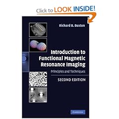
    Introduction to Functional Magnetic Resonance Imaging: Principles and Techniques 2nd edition

    Introduction to Functional Magnetic Resonance Imaging: Principles and Techniques 2nd edition by Richard B. Buxton provides an invaluable guide to how fMRI works, from introducing the basic ideas and principles to the underlying physics and physiology. He covers the relationship between fMRI and other imaging techniques and includes a guide to the statistical analysis of fMRI data. This book will be useful both to the experienced radiographer, and the clinician or researcher with no previous knowledge of the technology.
    Functional Magnetic Resonance Imaging (fMRI) has become a standard tool for mapping the working brain's activation patterns, both in health and in disease. In this second edition, Richard Buxton provides an invaluable guide to how fMRI works, from introducing the basic ideas and principles to the underlying physics and physiology.

    About the Author
    Richard B. Buxton is Professor of Radiology and Director of Magnetic Resonance Research at the University of California at San Diego.

    Book Details
    • Hardcover: 470 pages
    • Publisher: Cambridge University Press; 2 edition (October 5, 2009)
    • Language: English
    • ISBN-10: 0521899958
    • ISBN-13: 978-0521899956
    • Product Dimensions: 9.4 x 6.5 x 1.1 inches
    • List Price: $200.00
    • Price: $160.00
    • You Save: $40.00 (20%)
    Buy Now

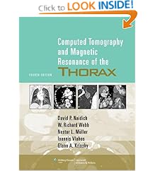
    Computed Tomography & Magnetic Resonance of the Thorax 4th Edition

    Computed Tomography & Magnetic Resonance of the Thorax 4th Edition by David P. Naidich, W. Richard Webb, Nestor L. Müller, Ioannis Vlahos & Glenn A. Krinsky provides authoritative, current guidelines on chest imaging using state-of-the-art technologies, including multidetector CT, MRI, PET, and integrated CT-PET scanning. This edition features a brand-new chapter on cardiac imaging. Extensive descriptions of the use of PET have been added to the chapters on lung cancer, focal lung disease, and the pleura, chest wall, and diaphragm. Also included are recent PIOPED II findings on the role of CT angiography and CT venography in detecting pulmonary embolism. Complementing the text are 2,300 CT, MR, and PET scans made on the latest-generation scanners.

    Book Details Computed Tomography & Magnetic Resonance of the Thorax
    • Hardcover: 897 pages
    • Publisher: Lippincott Williams & Wilkins; Fourth Edition (February 1, 2007)
    • Language: English
    • ISBN-10: 0781757657
    • ISBN-13: 9780781757652
    • Product Dimensions: 10.9 x 8.4 x 1.6 inches
    • List Price: $159.00
    • Price: $127.20
    • You Save: $31.80 (20%)
    Buy Now

    Copyright 2010 Radiology Book Review - All Rights Reserved.
    Designed by Web2feel.com | Bloggerized by Lasantha - Premiumbloggertemplates.com | Affordable HTML Templates from Herotemplates.com.