Showing posts with label Computed Tomography. Show all posts

Budoff: Handbook of Cardiovascular CT: Essentials for Clinical Practice

Handbook of Cardiovascular CT: Essentials for Clinical Practice has been created as a primer to help cardiologists, radiologists, and other cardiac-imaging enthusiasts to have a handy reference tool for the answers to important questions related to cardiovascular computed tomography (CCT). CCT has evolved from novel technology to research tool to essential clinical imaging modality at an astounding pace. The technology has great relevance for the multitude of medical and surgical disciplines focused on the treatment of cardiovascular disease.
Imaging of anatomy, physiology, and tissue characteristics from large-to-small vessel within seconds, and reconstruction to multimodal 2- and 3-D images within minutes, has facilitated practical clinical applications important to cardiovascular diagnosis, risk stratification, and procedure guidance. The new wave in technology requires education and training of initial generations of readers and users of CCT focused on providing an understanding of the essentials of methodology, technique, and image analysis for clinical application.

Ellis: Human Sectional Anatomy: Atlas of Body Sections, CT and MRI Images 3rd edition

Successive authors of atlases on sectional anatomy have emphasized the value to the anatomist and the surgeon of being able to view the body in this dimension. It is always difficult to consider three dimensions in the mind’s eye; to be able to view the relationships of the viscera and fascial planes in transverse and vertical section helps to clarify the conventional appearances of the body’s structure as seen in the operating theatre, in the dissecting room and in the textbook.
The introduction of modern imaging techniques, especially ultrasound, computed tomography (CT) and magnetic resonance imaging (MRI), has enormously expanded the already considerable importance of sectional anatomy. The radiologist, neurologist, internist, chest physician and oncologist, as well as specialists in the various fields of surgery, have had to re-educate themselves in the appearances and relationships of anatomical structures in transverse and vertical section. Indeed, precise diagnosis, as well as the detailed planning of therapy (for example, the ablative surgery of extensive cancer) and of interventional radiology, often depends on the cross-sectional anatomical approach.
This atlas combines three presentations of crosssectional anatomy – that of the dissecting room, CT and MRI. The series are matched to each other as closely as possible on opposite pages. Students of
anatomy, surgeons, clinicians and radiologists should find the illustrations of anatomical cross-sections (obtained by the most modern techniques of preparation and photographic reproduction) and the equivalent cuts on imaging (obtained on state-of-the art apparatus) both interesting and rewarding.

Kramer: Positron Emission Tomography Computed Tomography: A Disease-Oriented Approach

PET/CT epitomizes a marriage made in imaging heaven. The ability of PET to image cellular metabolism combined with the anatomic detail of CT represents a transformational approach to imaging neoplasia and other diseases. The task of PET/CT image interpretation is also challenging. This stems from the need to be highly skilled in both nuclear medicine and body/brain imaging. The impact of this technology hasbeen enormous in cancer, Alzheimer’s disease, and epilepsy; now representing an integral component of the standard workup and management of these patients.
This book aims to provide a thorough understanding of the technical demands involved in combining CT and FDG PET: patient preparation, acquisition techniques including potential pitfalls and limitations, and the basics of instrumentation and physics needed for developing cogent technical approaches. Technical advances and controversies including the use of CT contrast and, when clinically relevant, newer radiopharmaceuticals (beyond the most clinically available 18F-2-deoxy-fluoro-D-glucose or FDG) are addressed briefly.

PET-CT Beyond FDG: A Quick Guide to Image Interpretation

At present, more than 95% of PET studies worldwide are performed in oncologic patients, using F-18 Fluorodeoxyglucose (FDG). But, despite its high diagnostic accuracy in determining the pivotal role in the restaging (and staging) of the neoplasm, FDG is handicapped by false negative and positive results, creating limitations in the differential diagnosis of cancer. Moreover, PET-FDG shares with all the other diagnostic techniques the inability to answer all the questions of the oncologist, the surgeon, and the radiotherapist. It cannot function alone, either in the diagnostic field, or in giving all the information connected with prognosis and pursue a “tailored strategy” for each patient. Therefore, despite its primary role, there is a wide range of indications in oncologic patients that other radiotracers may be useful.
Although [18F]fluorodeoxyglucose (FDG) generally shows an excellent performance as a cancer-imaging agent when using PET-CT, there are some settings in which other radiopharmaceuticals offer advantages. Such non-FDG tracers are now gaining widespread acceptance not only in research but also in clinical practice.

Brain CT Scans in Clinical Practice

Across emergency rooms all over the world, thousands of patients are referred for brain CT scans daily. A radiologist often has to interpret the scan or a consultation has to be made to a neurosurgeon to review the scan. Most of this happens late at night and is a significant source of discontent. Thus having frontline physicians to be proficient in interpreting the emergency brain CT scan improves the efficiency of the whole pathway of care and is potentially life saving as time is of the essence for many patients with severe brain injury or stroke.
Interpretation of the emergency CT brain scan is a visual art. Comparison is made between the image in front of you and a reference image. For the experienced person, this reference image is imprinted in the mind, therefore comparison is quick. For the beginner, you can either carry several examples of every possible appearance of normal and abnormal scans to compare with or read this book! This book contains a few proven ways of quickly learning to interpret a brain CT scan, irrespective of your previous experience.
The radiologist’s experience is related to the number of hours he or she has spent looking at CT scans. The radiologist conveys his evaluation of the CT scan in words that often come in a particular sequence and combination. This book is about helping you to rapidly understand and confidently use the same language used by the radiologist.

CT Glossary

ALARA (As Low As Reasonably Achievable) : A concept to keep the radiation dose low but one should make sure the test remains diagnostic.

Attenuation : Brightness of an object, based upon density. Typically measured by use of the Hounsfield unit (HU), which is a scale developed by Sir Godfrey Hounsfield that attributes brightness of water as a HU of 0, and less dense objects being negative (i.e., air is typically −1000 HU), and more dense objects being positive (i.e., metal is typically +1000 HU).

Beam Hardening : An artifact with effects on image accuracy primarily due to high–density metal or calcium situated near a low-density structure (i.e., soft-tissue or lumen), which attenuates the X-ray signal from disproportionately low-energy X-rays.

Blooming An artifact on CT which is caused by severe calcification or metal, which causes the bright object to appear larger than it is (due to partial voluming).

Collimation : The amount of tissue imaged on one rotation of the detectors, calculated as the number of detectors multiplied by the slice thickness (i.e., 64 detectors at 0.625 = 40 mm).

Collimator : Shapes the X-ray beam. This controls the width, detector configuration, quality, and position of the X-ray beam.

Computed Tomography of the Lung - A Pattern Approach by J. A. Verschakelen (Springer) 2007

Most radiologists are involved on a daily basis in the performance and interpretation of CT studies of the lung, one of the most frequent radiological examinations in routine practice.
The currently available state-of-the-art MDCT technology enables us to study and display lung anatomy and gross pathology up to the level of the secondary pulmonary nodule in an exquisite way.
Prof. J.A. Verschakelen and Dr. W. De Wever have taken an original but highly didactic approach to explaining and teaching the CT features of lung diseases and other diseases with a pulmonary component.
The numerous superb colour drawings, together with the well chosen high resolution CT images, enable the reader to better understand the CT changes in patients with pulmonary
diseases, to recognise characteristic appearances and distribution patterns of the lung parenchyma, and finally how to use these patterns to make a correct diagnosis or to narrow down the number of differential diagnostic possibilities. The fact that all chapters of this book have been authored by the two editors only ensures that the specific concept and approach of this book is well respected throughout the whole volume.
I congratulate the authors, both internationally well known chest radiologists, for writing this excellent and remarkable work. I can strongly recommend it to all trainees in radiology and pneumology, as it provides a perfect guide through the field of HRCT of the lung, as well as to certified medical specialists who want to update their knowledge in CT of the lung.
I am confident that this volume will meet with the same success among readers as many of the previous volumes published in this series.

- Foreword from Albert L. Baert -

Contents
1 Introduction
2 Basic Anatomy and CT of the Normal Lung
3 How to Approach a CT of the Lung?
4 Increased Lung Attenuation
5 Decreased Lung Attenuation
6 Nodular Pattern
7 Linear Pattern
8 Case Study
Subject Index

About the Editor
J. A. Verschakelen, MD, Professor
W. De Wever, MD
Department of Radiology
University Hospitals Leuven
Herestraat, 49
3000 Leuven
Belgium

CT Evaluation of Coronary Artery Disease by Paolo Pavone (Springer) 2009

Coronary CT angiography (CTA) is rapidly changing the patient-care algorithms used to detect coronary artery disease, as well as the approach we take in risk-factor assessment and in the triage of patients. The rapid adoption of coronary CTA into clinical practice has been fueled by significant yearly advances in CT technology, which have improved the spatial and temporal resolution of this technique while simultaneously decreasing radiation exposure.
The growing utilization of coronary CTA has created a need for comprehensive didactic texts that explain the numerous applications of this new technology with respect to the pathophysiology of coronary artery disease, while also providing information on the approach to patients who have undergone previous bypass surgery or percutaneous coronary intervention. This book accomplishes both of these goals, and does so in a reader-friendly format. The image quality of the many figures that accompany each chapter is excellent and reflects the use of state of the art technology. The techniques described for plaque detection and characterization represent the current thinking pervasive in the coronary CTA community. The comprehensive reference list at the end of the book offers the reader a wealth of resources for further study.
There is no doubt that this book will be popular with radiologists, cardiologists, CT technologists and anyone else seeking to acquire a comprehensive understanding of coronary artery disease and its depiction using coronary CTA.

Contents
1 Clinical Anatomy of the Coronary Circulation
  • Angiographic Anatomy of the Coronary Circulation
  • Intramyocardial Vascularization and the Venous Circulation
  • Variability of the Coronary Artery Circulation
  • Anomalous Coronary Arteries
  • Factors Determining Coronary Artery Size
2 Basic Techniques in the Acquisition of Cardiac Images with CT
  • Technical Principles in the Acquisition of Cardiac Images by CT
  • From Conventional to Spiral CT
  • From Spiral to Multislice CT
  • Detector Number and Cardiac Imaging
  • Temporal Resolution in Cardiac Imaging
  • Types of Equipment and Their Clinical Uses in Cardiac Imaging
  • Other Factors That Improve the Image Quality of CT Technology
3 CT Examination of the Coronary Arteries
  • Achieving Excellent Image Quality in CT of the Coronary Arteries
  • CT Angiography of the Coronary Arteries
4 Image Reconstruction
  • Planimetric Techniques
  • Volumetric Techniques (Volume Rendering)
  • Virtual Endoscopy
5 Coronary Pathophysiology
  • Coronary Flow Reserve
  • Coronary Stenosis: Definition and Evaluation in Coronary Artery Disease
  • The Limits of Coronary Angiography
6 The Atherosclerotic Plaque
  • The Vulnerable Plaque: Biology and Histology
  • The Vulnerable Plaque: Local and Systemic Factors Contributing to Plaque Rupture
7 Intravascular Ultrasound: From Gray-Scale to Virtual Histology
  • Introduction
  • From Gray-Scale to Color-Coded IVUS: The Virtual-Histology Revolution
  • Lesion Classification Using IVUS-VH
  • IVUS-VH Console and Image Interpretation: Tips and Tricks
8 Identification and Characterization of the Atherosclerotic Plaque Using Coronary CT Angiography
  • Normal Vascular Wall
  • Identification of Atherosclerotic Plaques in Coronary CT Angiography
  • CT Density Values and Plaque Characterization: Fibrolipidic and Calcific Plaques
  • Atherosclerotic Plaque and Disease Evolution
  • Diagnostic Evaluation of Coronary Disease During Medical Therapy
9 Coronary CT Angiography: Evaluation of Stenosis and Occlusion
  • Non-Significant Moderate Stenosis
  • Calcified Plaques: Problems in Defining Vascular Stenosis
  • Significant Stenosis
  • Remodeling
  • Occlusion of the Coronary Arteries and the Development of Collateral Circulatio
  • Evaluation of Coronary-Artery Stenosis: A Review of the Literature
  • Saving Lives
10 Current Strategies in Cardiac Surgery
  • Standard Grafting Techniques
  • Results
11 Coronary CT Angiography: Evaluation of Coronary Artery Bypass Grafts
  • Pre-Operative CT Evaluation
  • Post-Operative Evaluation of CABG
  • CT Evaluation of CABG: Technique
  • CT Evaluation of CABG: Results
12 Coronary Stents
  • Types of Stents
  • Mechanism of Stent Expansion
  • Materials
  • Fabrication Methods
  • Additions
  • Impact of Stent Design on Clinical Outcome
  • Bioabsorbable and Biocompatible Stents
13 CT Angiography of Coronary Stents
14 X-ray Exposure in Coronary CT Angiography
  • Damage from Ionizing Radiation
  • X-ray Dose During CT
  • Techniques for Limiting X-ray Exposure in Coronary CT Angiography
  • X-ray Exposure and Patient Age
15 Use of MSCT Scanning in the Emergency-Room Evaluation of Patients with Chest Pain
  • Causes of Acute Chest Pain
  • Multislice CT Scanning in Acute Chest Pain
  • The “Triple Rule Out” Protocol
16 Current Recommendations for Coronary CT Angiography
  • Technical Considerations
  • Evaluation of Coronary Stenoses in Patients at Low or Intermediate Risk of Cardiovascular Disease
  • Evaluation of Coronary-Artery Bypass Patency
  • Other Frequent Uses of Coronary CT Angiography
  • Contraindications to Coronary CT Angiography
  • Future Directions in Non-invasive Coronary Artery Imaging with Coronary CT Angiography
17 Prognostic Value of Coronary CT
Suggested Readings

Originally published as:
Malattia coronarica
Fisiopatologia e diagnostica non invasiva con TC
Paolo Pavone, Massimo Fioranelli
© Springer-Verlag Italia 2008
All rights reserved


Le malattie cardiovascolari rappresentano attualmente la causa principale di mortalità e, oltre a modificare sensibilmente la qualità della vita, comportano un notevole impegno economico per la società. Poiché la maggior parte degli eventi coronarici si verifica per la complicanza di una placca aterosclerotica parietale non stenosante, il suo riconoscimento può assumere rilevante significato clinico ed essere di interesse nella scelta di un trattamento medico o interventistico.
Attraverso l’utilizzo di apparecchiature sempre più sensibili e veloci, oggi la TC coronarica ci consente, finalmente, di visualizzare la lesione responsabile delle sindromi coronariche acute e di caratterizzarla. La conoscenza delle apparecchiature, la valutazione dei loro limiti e l’adeguata preparazione del paziente, rappresentano passaggi importanti per l’ottenimento di immagini adeguate dal punto di vista diagnostico.
L’opera nasce dalla volontà di fornire al cardiologo o al medico non esperto di imaging le basi per comprendere principi tecnici e modalità di acquisizione e di ricostruzione delle immagini. Allo stesso tempo anche il radiologo che non abbia esperienze specifiche di imaging cardiaco potrà acquisire conoscenze di base di anatomia e fisiopatologia delle coronarie.

Cardiac CT by Marc Dewey (Springer) 2011

Computed tomography has been going through a dramatic evolution of technology in the past years. The increased spatial and temporal resolution directly translate into improved image quality and more versatile applications. Indeed, entirely new areas of clinical application have emerged, one of the most prominent being the recent advent of cardiac computed tomography. Especially CT angiography of the coronary arteries – often referred to as coronary CT angiography – has received tremendous interest and is currently entering the clinical arena.
In fact, it has the potential to greatly alter the way in which many patients with suspected coronary artery disease will be worked up. However, the technique is still relatively new to the medical community. Furthermore, in the recent past several developments – such as substantial further improvement of scanner technology and the extensive use of low-dose acquisition protocols – have been introduced and will influence the utilization of cardiac CT. At this stage, it is crucial that all those potentially involved in the new imaging technology – those who perform and interpret the scan and also those who order a cardiac CT and advise their patients as to whether a CT examination might be the right test for them – are well informed about the technology and answers it can provide, about its limitations and problems, and about how to best apply it in a given clinical situation.
This is why the textbook edited by Dr. Dewey is most welcome. With ample illustrations, it provides the technological background and principles of scan acquisition and interpretation as well as outlines the range of clinical applications of cardiac CT. May it be a useful resource to its readers and may it contribute towards the further development of this exciting field.

Contents
1 Introduction
2 Technical and Personnel Requirements
3 Anatomy
4 CT in the Context of Cardiovascular Diagnosis and Management
5 Cardiac CT in Clinical Practice
6 Clinical Indications
7 Patient Preparation
8 Physics Background and Radiation Exposure
9 Examination and Reconstruction
10 Examinations on Different CT Scanners
10a Toshiba Aquilion 64 and Aquilion ONE
10b Siemens Somatom Sensation and Definition
10c Philips Brilliance 64 and iCT
10d General Electric Light Speed VCT and Discovery CT750HD
11 Reading and Reporting
12 Coronary Artery Bypass Grafts
13 Coronary Artery Stents
14 Coronary Artery Plaques
15 Cardiac Function
16 Cardiac Valves
17 Coronary Artery Anomalies
18 Congenital and Acquired Heart Disease
19 Typical Clinical Examples
20 Results of Clinical Studies
21 Outlook
Index

Kau: Cone Beam CT of the Head and Neck: An Anatomical Atlas

Cone Beam CT of the Head and Neck - An Anatomical Atlas is dedicated to the memory of the three anonymous individuals whose cadaver prosections are the subject matter of this atlas. They unknowingly have made a donation to science that will benefit students and clinicians in the imaging sciences. The handling of the cadaver donations that are displayed in this clinical atlas was managed with respect and driven by scientific yearn for discovery; the discovery to benefit the current and next generation of clinicians and researchers. These donations will enrich the basic foundational knowledge base of human anatomy as depicted in cone beam CT imaging. The current effort made it possible to correlate the CBCT images with the actual physical image. This is a foundational knowledge base from which to build and make new discoveries. Future generations can then build on this foundational knowledge base to identify bone density states, tissue function profiles (atrophy, hypertrophy, etc) disease states (neoplasia, metaplasia, etc.). We trust that the boundaries have few limits.
This pocket clinical atlas was produced to help dental and medical colleagues understand and correlate structures of the head and neck with cone beam computerized tomography (CBCT) imaging technology.
Methods used for radiographic evaluation and diagnosis have undergone enormous changes in the last 20 years. New technologies are being developed and are becoming readily available to the medical and dental field. The advancements in hardware and software have allowed the development of innovative methods for facial diagnosis, treatment planning, and clinical application.
CBCT was developed in the 1990s as an advancement in technology resulting from the demand for three-dimensional (3D) information obtained by conventional computed tomography (CT) scans. The development of CBCT technology reduces exposure by using lower radiation dose, compared with conventional CT. As the demand for the technology increases, so has the market for custom built craniomaxillofacial CBCT devices. The rates of increase for CBCTs have been increasing in number on the market over the last decade and a variety of applications to the facial and dental environments have been established.

Contents
  • Introduction
  • Conventional Computed Tomography
  • CBCT
  • CBCT Data
  • CBCT Acquisition Systems
  • Uses of CBCT Technology
  • Diagnosis
  • Clinical Applications of the CBCT
  • Late Evaluation with CBCT
  • The Purpose of the Clinical Atlas
  • References
  • Axial
  • Coronal
  • Sagittal 
Product Details 
  • Paperback: 75 pages
  • Publisher: Springer; 1st edition (March 9, 2011)
  • Language: English
  • ISBN-10: 3642127037
  • ISBN-13: 978-3642127038
  • Product Dimensions: 7.5 x 5 x 0.4 inches
 List Price: $69.95

    Multidetector CT in Neuroimaging - An Atlas and Practical Guide (Atlas Medical Publishing) 2009

    Multidetector computed tomography (MDCT) offers new and exciting opportunities for imaging patients suspected of afflictions of the nervous system. Achievement of this benefit requires an understanding of its full potential in displaying the features of the spectrum of relevant diseases. It also depends upon employing an approach that ensures that it is applied to best effect by tailoring the technique of examination to the patient's clinical problem.
    This book will provide practical guidance to radiologists, whether general or specialist, and their team of radiographers/technicians that will enable them to take full advantage of the opportunities offered by MDCT in brain and spine imaging. This will enrich their practice and expand the benefits they can bring to their patients.
    The advantages of MDCT include its ability for routine sub-millimetre scanning of large areas at acceptable radiation doses. The enhanced postprocessing techniques and the rapidity and ease with which they can be obtained mean that they can be applied with no limitation on throughput or reporting times. Although magnetic resonance imaging (MRI), with its ability to differentiate soft tissues has many applications, computed tomography (CT) remains the appropriate first-line investigation for most patients with an acute cerebral event and for those who still cannot undergo MR for one reason or another (approximately 1 patient in 5).
    MDCT has many technical benefits over single-slice CT but when a small area is to be covered, e.g. the orbit and the cavernous sinus, a single slice scanner is acceptable and a 4-slice MDCT can be almost as effective as a 64-slice CT. In contrast, when a large area is to be covered, a scanner with 16 or more detectors is optimal and single slice imaging precluded by technical limitations and the radiation dose it would give. MDCT provides new opportunities to improve the standard of routine brain examinations and to expand CT techniques into new areas. The techniques we describe can be adapted to any MDCT system for any similar clinical situation
    In most radiology textbooks a systematic approach is followed. The appearances of specific pathologies, e.g tumours, trauma, and degenerative disease, are classified, described and inclusively illustrated. In practice, most patients present with a combination of symptoms and signs which must be investigated in order that pathology can be either excluded or identified and managed. This book adopts such a problem-based approach and is intended to be a supplement to the standard disease-based texts. Our approach is based on the concept that the information from the clinical referral can be used to define a tailored MDCT technique which will optimize the likelihood of correctly identifying or excluding causative pathology. The commonly held belief that a single CT brain protocol is appropriate in all situations is no longer realistic, appropriate, or tenable.
    The illustrations are intended to convey how the CT techniques applied affected diagnosis or management in a range of common clinical scenarios. They show how MDCT can be used in a routine service role in a way that maximizes its potential, and how knowledge of that potential enables its use to be extended to unexpected applications. The illustrations are not inteded to be a comprehensive catalogue of abnormalities, but to convey why such images should be obtained and how to get the most from them. Protocols are given in detail so that radiographers or technicians, with the guidance of the radiologist, can apply them in any specialist or nonspecialist department.
    Detailed imaging of a patient suspected of disease of the brain or other parts of the nervous system need no longer be the prerogative of a specialist neuroradiology unit. Using the approaches we describe, detailed, highly focussed, highly informative and clinically influential investigation can be practised in any radiology department where there is a need and desire to provide quality brain and spinal CT imaging.
    This book will appeal to many kinds of reader and at different stages of their careers. It will be an invaluable companion to trainee and trained radiologists in their daily study and work. Radiographers/technicians will find the reasons that a specific protocol is chosen will enhance their knowledge and understanding and make their work more interesting and fulfilling. Clinicians will gain insight into the radiologists mind: how and why they make decisions and why specific techniques are chosen. Most of the common neurological and neurosurgical conditions are covered so that a review of the images will be a valuable exercise in preparation for the ordeals of post-graduate examinations!

    Contents
    1 Routine noncontrast brain imaging
    2 Contrast enhanced brain imaging
    3 Orbital pathology
    4 Visual failure
    5 Pituitary imaging
    6 Cranial nerve palsies 3–6
    7 Intracranial imaging in stroke: ischaemic
    8 Vascular imaging in ischaemic stroke and TIA
    9 Acute haemorrhagic stroke
    10 Variations in arterial anatomy
    11 Venous pathology and variations in venous anatomy
    12 Cranial nerve palsies 7–12
    13 Spinal imaging
    Concluding comments
    Index

    Anatomy of the Heart by Multislice Computed Tomography by Francesco Fulvio Faletra (Wiley-Blackwell) 2008

    Anatomy is perhaps the simplest of medical sciences, requiring little more than some curiosity plus careful observation of things as they are. The new multislice computed tomography (MSCT) machines produce a volume data set with the highest isotropic spatial resolution ever seen. The 0.6-mm “pixel” (picture element) that CT has traditionally delivered in axial planes (x and y dimensions) is also extended to the z dimension as well. The spatial resolution of these new machines is such that they can scan a 10-mm diameter piece of heart in 20 axial sections to produce nearly 4000 pixels for each slice. In other words, these scanners are capable of digitizing the anatomy of a 70-kg human body into over half a billion individual voxels (volume elements).
    Given this high spatial resolution, MSCT offers superb 3D images of the entire heart and great vessels. Relationships between cardiac structures can be shown as never before. Electronic casts and electronic dissections of the heart in any plane can show the internal and external cardiac structures.
    As a result, the anatomy of the heart and great vessels can be understood easily by young doctors, medical students and nurses. This atlas has been made for them.
    MSCT is unique in its ability to image coronary arteries and it is likely to become one of the most used cardiac imaging techniques. Images of coronary vessels provided by MSCT can be interpreted both from radiological and cardiological standpoints. Indeed, radiologists are often not sufficiently familiar with cardiac anatomy, whereas cardiologists often lack adequate familiarity with the axial, coronal or sagittal planes used for visualization of cardiac MSCT images. This atlas has also been made to clarify the anatomy for both specialties.
    The atlas is divided into 10 chapters. In each chapter, the body planes, cardiac planes and cardiac structures, such as cardiac chambers, cardiac valves, septa, coronary arteries and coronary veins, are displayed from many perspectives to give the reader a wider vision of living cardiac anatomy.

    Contents
    1 Basic Principles
    2 Location of the Heart: Body Planes and Axis
    3 Cardiac Planes
    4 The Right Heart
    5 The Left Heart
    6 The Cardiac Valves
    7 The Cardiac Septum
    8 Coronary Artery Anatomy
    9 Coronary Vein Anatomy
    10 Coronary Artery Bypass Grafts
    Index

    Revisiting Cardiac Anatomy - A Computed-Tomography-Based Atlas and Reference by Farhood Saremi (Wiley-Blackwell) 2011

    The past two decades have witnessed a revolution not only in management but also in the diagnostic approach to cardiovascular disease. Modern imaging tools provide invasive and noninvasive strategies that allow increasingly accurate characterization of morphology and function of the heart. Nuclear medicine, echocardiography (including transesophageal, intravascular, and intracardiac imaging) as well as cardiac magnetic resonance have all immensely contributed to the ability to provide accurate diagnostic and prognostic information. At the same time, modern surgical and interventional treatment requires accurate anatomic information and guidance. The success of increasingly complex coronary and noncoronary structural interventions as well as electrophysiological procedures depends on ever more detailed imaging support. The interface between imaging, diagnosis, and treatment is of tremendous importance and at the same time needs to face increasingly challenging requirements.
    In addition to the more established techniques, highresolution multidetector CT imaging of the heart and cardiovascular system has recently entered the stage. Modern hardware allows truly isotropic imaging with high temporal resolution and a spatial resolution of 0.5 mm or better. Two-dimensional and three-dimensional reconstructions allow to perform “virtual dissection” of complex cardiac anatomy with phenomenal image quality. In fact, in vivo imaging will often provide a more realistic depiction of true anatomic relationships than postmortem analysis.
    However, while modern imaging provides new possibilities, it also creates new needs and challenges. To fully realize the potential inherent to the large, detailed datasets, a new type of “imager,” needs to fulfill many roles—from teaching medical students and residents to helping the cardiologist better define and the interventionalist or surgeon better correct cardiovascular defects by fully understanding the underlying anatomy.
    Revisiting Cardiac Anatomy - A Computed-Tomography-Based Atlas and Reference by Farhood Saremi provide a valuable resource and useful companion to a broad spectrum of readership, from medical students starting their career in a dissection hall to cardiologists and cardiothoracic surgeons who rely on ever more accurate imaging for diagnosis and treatment. This atlas is a fresh approach to cardiac anatomy that provides high-quality in vivo anatomy through carefully rendered images with explanatory texts of the clinically relevant underlying anatomic concepts. In spite of amazing proliferation of information on the Internet and multiple Web sites filled with up-to-date information, there is no similarly detailed and systematic compilation of morphological imaging with CT to demonstrate normal in vivo anatomy of the heart and its variants. For good understanding of health and disease, a good anatomy book is irreplaceable and will allow both for systematic learning and as a quick, yet detailed reference for specific clinical questions.
    The fantastic imaging quality that can be achieved with state-of-the-art CT imaging hardware and protocols in the hands of experts has deeply impressed and convinced us. The author are confident that it will also convince our readership: imaging is a virtual reality.

    Contents
    • Chapter 1: Anatomy of the Heart for a Dissector
    • Chapter 2: Anatomical and Pathophysiological
    • Classification of Congenital Heart Disease
    • Chapter 3: CT in Pediatric Heart Disease
    • Chapter 4: Mitral and Aortic Valves Anatomy for Surgeons and Interventionalists
    • Chapter 5: Clinical Applications of CT Imaging of the Aortic and Mitral Valves
    • Chapter 6: Computed Tomography for Percutaneous Aortic Valve Replacement
    • Chapter 7: Mitral Valve Disease Imaging
    • Chapter 8: The Aortic Root
    • Chapter 9: CoronaryAnatomyforInterventionalists
    • Chapter 10: Coronary Anatomy for Surgeons
    • Chapter 11: Anatomy for Electrophysiologic Interventions
    • Chapter 12: Coronary Atherosclerosis: CT Imaging for the Preventive Cardiologist
    • Chapter 13: Nomograms for Coronary Computed Tomographic Angiography
    • Appendix
    • Index

    CT and MR Angiography of the Peripheral Circulation - Practical Approach with Clinical Protocols by Debabrata Mukherjee (Informa Healthcare) 2007

    The advent of multislice computed tomographic angiography (CTA) and magnetic resonance angiography (MRA) has had a dramatic impact on practice patterns in cardiovascular medicine with a significant proportion of catheterbased diagnostic angiography giving way to these new modalities.
    This text represents a comprehensive treatise on CTA and MRA for the diagnosis of peripheral vascular disease and represents a consolidated attempt at bridging the everwidening gap between innovation in these areas and the knowledge of the general and cardiovascular clinician. A large number of high-quality illustrations make this textbook particularly attractive to the general clinician. Abundant tables and scanning protocols are included in each chapter wherever applicable.
    In the first half of the book dedicated to CTA, the introductory chapters address technical principles, image reconstruction, radiation dosimetry, and principles of CT contrast agents and their administration. Subsequently, clinical application of CTA in each major vascular territory is dealt with thoroughly, with clear description of the examination and scanning technique for assessing each specific territory. The section on MRA provides a state of the art overview of this methodology in the peripheral circulation. MR physics relevant to the clinician is succinctly articulated with specific chapters dedicated to pulse sequences commonly used in peripheral angiography and MR contrast agents. Individual chapters on each major vascular territory including the extracranial and intracranial circulation, the pulmonary circulation, the thoracic and the abdominal aorta, the renal, and mesenteric circulation, the lower and upper extremity circulation enable the reader to appreciate the intricacies of MRA techniques for the evaluation of these territories.
    In summary, Doctors Debabrata Mukherjee, Sanjay Rajagopalan, Informa Healthcare and colleagues have put together a very didactic and practical book. I believe the information will serve to stimulate the interest not only of the cardiovascular specialist ready to become familiarized with this important technological development of CT and MRI as applied to patients with peripheral vascular disease, but also of a broad range of professionals and students of cardiovascular medicine.
    This book is a valuable resource and should help the efforts to incorporate CT and MR in mainstream cardiovascular practice.
    Valentin Fuster MD PhD
    Professor of Medicine
    Director, Mount Sinai Heart
    Mount Sinai Medical Center
    New York City, NY

    Contents
    Section 1: CT angiography

    1. Fundamentals of multi-slice CT scanning and its application to the periphery
    2. Image reconstruction with multi-slice CT
    3. Radiation dosimetric considerations in peripheral CT angiography
    4. Image post-processing in CT
    5. X-ray contrast agents and contrast timing considerations
    6. CT angiography of the extracranial and intracranial circulation with imaging protocols
    7. CT angiography and venography of the pulmonary circulation with imaging protocols
    8. CT angiography of the thoracic aorta with protocols
    9. CT angiography of the abdominal aorta and its branches with protocols
    10. CT angiography of the upper extremities with protocols
    11. CT angiography of the lower extremity circulation with protocols
    Section 2: MR Angiography
    12. Fundamental principles of MR imaging
    13. MR techniques for vascular imaging
    14. Contrast agents in MR angiography
    15. MR angiography of the extracranial and intracranial circulation with imaging protocols
    16. MR angiography of the pulmonary circulation
    17. MR angiography of the thoracic aorta with protocols
    18. MR angiography of the abdominal aorta with protocols
    19. MR angiography of the renal circulation with imaging protocols
    20. MR angiography of the mesenteric circulation with imaging protocols
    21. MR angiography of the lower extremity circulation with protocols
    22. MR venography: practical imaging techniques
    Index

    Rubin: CT and MR Angiography: Comprehensive Vascular Assessment

    Written by world-renowned experts in both CT angiography and MR angiography, this landmark work is the first comprehensive text on vascular imaging using CT and MR. It provides a balanced view of the capabilities of these modalities and practical guidelines for obtaining and interpreting images. More than 2,200 illustrations complement the text. Chapters co-authored by CT and MR authorities cover imaging of all coronary and non-coronary arteries and veins. Each chapter details indications, imaging strategies, normal and variant anatomy, diseases, surgical management, and pitfalls. The authors compare the utility of CT and MR in specific clinical situations and discuss the role of conventional angiography and ultrasound where appropriate.

    Contents 
    Part I. IMAGING PRINCIPLES
    • Principles of Computed Tomographic Angiography
    • Principles of Magnetic Resonance Angiography
    • Conventional Angiography in the Noninvasive Era
    • Contrast Medium Administration: Computed Tomographic Angiography
    • Contrast Administration: Magnetic Resonance Angiography
    • Postprocessing and Data Analysis
    Part II. ESSENTIALS OF VASCULAR DISEASE
    • Atherosclerosis: Epidemiology and Pathophysiology
    • Atherosclerosis:In Vivo Characterization
    • Thromboembolism
    • Vasculitides and Connective Tissue Disorders
    Part III. VASCULAR ANATOMY AND PATHOLOGY
    • Cerebral Arteries and Veins
    • Cervical Vasculature
    • Quantifying Coronary Artery Calcium
    • Coronary Arteries
    • Thoracic Vascular Anomalies
    • Pulmonary Vasculature
    • Thoracic Aorta
    • Abdominal Aorta
    • Mesenteric Vasculature
    • Renal Vasculature
    • Lower Extremities Vasculature
    • Upper Extremities Vasculature
    • Pediatric Techniques and Vascular Anomalies
    • Venous System


    Book Details
    • Hardcover: 1316 pages
    • Publisher: Lippincott Williams and Wilkins Publishers; 1 edition (August 1, 2008)
    • Language: English
    • ISBN-10: 078174525X
    • ISBN-13: 978-0781745253
    • Product Dimensions: 11.1 x 9 x 2.2 inches
    List Price: $269.00

    High-Resolution CT of the Lung 4th Edition

    High-Resolution CT of the Lung 4th Edition by W. Richard Webb, Nestor L. Müller & David P. Naidich explains how to use the newest high-resolution CT technology to detect and diagnose lung abnormalities. Still the only complete text on the topic, this compact, affordable reference is written by the foremost experts and provides cutting-edge technical and clinical information. It includes state-of-the-art HRCT scans of interstitial lung diseases and differential diagnosis tables summarizing the most helpful diagnostic features of interstitial and airspace diseases.
    This edition includes full-color illustrations of histologic findings in lung disease, correlated with HRCT manifestations. Also included are updated HRCT images obtained on multidetector CT scanners with many coronal and sagittal reformations. Two new chapters on the idiopathic interstitial pneumonias detail the differential diagnosis, pathophysiology, histology, clinical manifestations, and HRCT features of these entities. A companion Website will offer the fully searchable text plus an image bank containing all illustrations from the text.

    Book Details High-Resolution CT of the Lung
    • Hardcover: 672 pages
    • Publisher: Lippincott Williams & Wilkins; Fourth Edition (October 1, 2008)
    • Language: English
    • ISBN-10: 0781769094
    • ISBN-13: 9780781769099
    • List Price: $210.00
    Buy Now

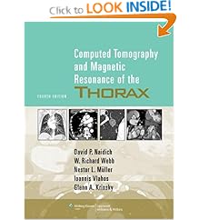
    Computed Tomography & Magnetic Resonance of the Thorax 4th Edition

    Computed Tomography & Magnetic Resonance of the Thorax 4th Edition by David P. Naidich, W. Richard Webb, Nestor L. Müller, Ioannis Vlahos & Glenn A. Krinsky provides authoritative, current guidelines on chest imaging using state-of-the-art technologies, including multidetector CT, MRI, PET, and integrated CT-PET scanning. This edition features a brand-new chapter on cardiac imaging. Extensive descriptions of the use of PET have been added to the chapters on lung cancer, focal lung disease, and the pleura, chest wall, and diaphragm. Also included are recent PIOPED II findings on the role of CT angiography and CT venography in detecting pulmonary embolism. Complementing the text are 2,300 CT, MR, and PET scans made on the latest-generation scanners.

    Book Details Computed Tomography & Magnetic Resonance of the Thorax
    • Hardcover: 897 pages
    • Publisher: Lippincott Williams & Wilkins; Fourth Edition (February 1, 2007)
    • Language: English
    • ISBN-10: 0781757657
    • ISBN-13: 9780781757652
    • Product Dimensions: 10.9 x 8.4 x 1.6 inches
    • List Price: $159.00
    • Price: $127.20
    • You Save: $31.80 (20%)
    Buy Now

    Fundamentals of Body Ct 3rd Edition

    Fundamentals of Body Ct 3rd Edition by W. Richard Webb, William Brant and Nancy Major makes it remarkably simple to learn how to perform and interpret CT scans. The completely revised and updated 3rd Edition covers the most recent advances in CT technique, including the use of multislice CT to diagnose chest, abdominal, and musculoskeletal abnormalities, as well as the expanded role of 3D CT and CT angiography in clinical practice. The result is today's most accessible, affordable introduction to body CT!

    Book features:
    • Highlights the information essential for interpreting CTs and the salient points needed to make diagnoses.
    • Reviews how the anatomy of every body area appears on a CT scan.
    • Presents comprehensive guidance in a concise format.
    • Offers step-by-step instructions on how to perform all current CT techniques.
    • Provides a survey of major CT findings for a variety of common diseases-with an emphasis on those findings that help to differentiate one condition from another.
    New in this edition:
    • Features new chapters on CT of abdominal and pelvic trauma.
    • Presents updated abdominal chapters with recently reported findings and the latest pathological terminology.
    • Offers a revised musculoskeletal section that highlights abnormalities of the axial skeleton typically identified or diagnosed using body CT.
    • Includes increased coverage of high-resolution lung CT and solitary nodules.
    • Provides full-chapter coverage of hot topics such as (Ch. 4) Mediastinum - Lymph Node Abnormalities & Masses • (Ch. 6) Lung Disease • (Ch. 18) Trauma CT of the Abdomen & Pelvis • and (Ch. 19) CT Angiography of the Abdomen & Pelvis.
    About the Author
    W. Richard Webb, MD, Professor of Radiology, Chief, Thoracic Imaging, University of California, San Francisco, San Francisco, CA, USA; Wiliam E. Brant, MD, Professor of Clinical Radiology, Division Head of Thoraco-Abdominal Imaging, University of Virginia, Charlottesville, VA, USA; and Nancy Major, MD, Associate Professor of Radiology, Division of Skeletal Radiology, Duke University School of Medicine, Durham, NC.

    Book Details Body Ct
    • Paperback: 429 pages
    • Publisher: Saunders; 3 edition (October 3, 2005)
    • Language: English
    • ISBN-10: 1416000305
    • ISBN-13: 9781416000303
    • Product Dimensions: 10.2 x 7.2 x 0.8 inches
    • List Price: $79.00
    • Price: $64.53
    • You Save: $14.47 (18%)
    Buy Button

    Mettler Essentials of Nuclear Medicine Imaging 5th edition

    • Ch. 1 Radioactivity, Radionuclides, and Radiopharmaceuticals.
    • Ch. 2 Instrumentation.
    • Ch. 3 Quality Control.
    • Ch. 4 Cerebrovascular System.
    • Ch. 5 Thyroid, Parathyroid, and Salivary Glands.
    • Ch. 6 Cardiovascular System.
    • Ch. 7 Respiratory System.
    • Ch. 8 Gastrointestinal Tract. .
    • Ch. 9 Skeletal System.
    • Ch. 10 Genitourinary System and Adrenal Glands.
    • Ch. 11 Conventional Neoplasm Imaging and Radioimmunotherapy.
    • Ch. 12 Genitourinary System.
    • Ch. 13 Inflammation and Infection Imaging.
    • Ch. 14 Legal Requirements and Radiation Safety.
    • Self Evaluation Section.
    • Unknown Case Sets.
    • Answers to Unknown Case Sets.
    Appendices.
    • A. Characteristics of Radionuclides for Imaging Therapy.
    • B-1. Radioactivity Conversion Table for International System (SI) Units (Becquerels to Curies).
    • B-2. Radioactivity Conversion Table for International System (SI) Units (Curies to Becquerels).
    • C-1. Technetium 99m Decay and Generation Tables.
    • C-2. Iodine 131 and Strontium 89 Decay Charts.
    • D. Injection Techniques and Pediatric Dosage.
    • E. Sample Techniques for Nuclear Imaging.
    • F. Non-radioactive Pharmaceuticals in Nuclear Medicine.
    • G. Pregnancy and Breastfeeding.
    • H-1. General Considerations for Patients Receiving Radionuclide Therapy.
    • H-2. Special Considerations and Requirements for Iodine 131 Therapy.
    • I. Emergency Procedures for Radioactive Spills.
    Index.


    Essentials of Nuclear Medicine Imaging 5th edition by Fred A. Mettler and Milton J. Guiberteau has established itself as the best introduction to nuclear imaging techniques. It is practical, yet comprehensive, covering physics, instrumentation, quality control, and legal requirements. The 5th Edition features a new color format, with many user-friendly features such as "Pearls and Pitfalls." More than 600 pictures in digital-quality resolution depict imaging of each body system. A series of Unknown Case Sets, with answers, help test your knowledge.


    Book features:
    • Includes helpful appendices including Injection Techniques, Pediatric Dosages, Non-radioactive Pharmaceuticals, and many more.
    • Presents important "Pearls and Pitfalls" in each chapter.


    New in this edition:
    • Features a new full-color format making information easy to read and find.
    • Covers new techniques such as PET/CT, cardiac-gated SPECT, and tumor-specific radionuclides.
    • Provides full-chapter coverage of hot topics such as:
      • Cerebrovascular System.
      • Cardiovascular System.
      • Conventional Neoplasm Imaging and Radioimmunotherapy.
      • Positron Emission Tomography Imaging.
    • Includes seven complete Unknown Case Sets for self-testing.


    About the Authors
    • Fred A. Mettler, Jr., MD, MPH, Professor and Chair, Department of Radiology and Nuclear Medicine, University of New Mexico Health Sciences Center, Albuquerque, NM. 
    • Milton J. Guiberteau, MD, Professor of Clinical Radiology, University of Texas Medical School at Houston; Chairman, Department of Radiology, St. Joseph's Hospital, Houston, TX.


    Book Details

    • Hardcover: 592 pages
    • Publisher: Saunders; 5 edition (November 23, 2005)
    • Language: English
    • ISBN-10: 0721602010
    • ISBN-13: 9780721602011
    • Product Dimensions: 10 x 8 x 0.8 inches
    List Price: $106.00 
     

    Copyright 2010 Radiology Book Review - All Rights Reserved.
    Designed by Web2feel.com | Bloggerized by Lasantha - Premiumbloggertemplates.com | Affordable HTML Templates from Herotemplates.com.