Showing posts with label Gastrointestinal Imaging. Show all posts

Cotton: Advanced Digestive Endoscopy: Practice & Safety

  • Chapter 1: Overview.
  • Chapter 2: Design and Management of Gastrointestinal Endoscopy Units.
  • Chapter 3: Sedation, Analgesia, and Monitoring for Endoscopy.
  • Chapter 4: Endoscopic Equipment.
  • Chapter 5: Digital Documentation in Endoscopy.
  • Chapter 6: Principles of Electrosurgery, Laser and Argon Plasma Coagulation with Particular Regard to Colonoscopy.
  • Chapter 7: Infection Control in Endoscopy.
  • Chapter 8: Risks, Prevention and Management.
  • Chapter 9: Pathology.
  • Chapter 10: Pediatric Gastrointestinal Endoscopy.
  • Chapter 11: Training and Credentialing in Gastrointestinal Endoscopy.
  • Chapter 12: Towards Excellence and Accountability.


Endoscopy is a technically challenging procedure. Until recently it was regarded as a routine procedure that could be performed by general gastroenterologists until outcome and follow-up analyses revealed unacceptably high levels of complications. The opinion-leaders in endoscopy have brought about a shift in attitude. The emphasis is now very much on a 'back to basics' approach with retraining recommended for all levels of doctors performing endoscopy - and several techniques restricted to only the very experienced.

This series is aimed at the endoscopist with some experience who wishes to achieve competency in the more advanced techniques.

The book includes information and guidelines for all aspects of practice management relating to endoscopy. It provides a practical manual on how to perform techniques in the most safe and effective ways to reduce complications.

It covers training, endoscopy and imaging equipment, set-up of endoscopy units, cleaning & disinfection and patient preparation and monitoring. It also contains techniques for screening, diagnosis, treatment and follow-up from THE leading international names.


Key Features
  • Provides a practical manual on how to perform techniques safely and effectively in order to maximise value, and to reduce risks.
  • Covers training, endoscopy and imaging equipment, infection control, patient preparation and monitoring, complications and how to avoid and deal with them.
  • Contains techniques for screening, diagnosis, treatment and follow-up from THE leading international names.
  • Includes information and guidelines for all aspects of practice management relating to endoscopy.


About the Author
  • Peter Cotton was born in England, educated at Cambridge University and at St. Thomas Hospital Medical School (London). He became interested in endoscopy in the late 1960’s with the introduction of flexible fiberscopes, and developed units at St. Thomas’ Hospital and at the Middlesex Hospital, which pioneered and evaluated many diagnostic and therapeutic procedures, particularly ERCP. He attracted postgraduates from many countries, held numerous teaching courses, and introduced live CCTV workshops. In 1986 he became Professor of Medicine and Chief of Endoscopy at Duke University in North Carolina, and developed a state of the art endoscopy center. He maintained his interests in teaching, new techniques, and careful outcome evaluation. He moved to Charleston, South Carolina in 1994 to initiate and lead a Digestive Disease Center, dedicated to multi-disciplinary patient care, and the research and education needed to enhance it.


Book Details

  • Hardcover: 384 pages
  • Publisher: Wiley-Blackwell; 1 edition (May 6, 2009)
  • Language: English
  • ISBN-10: 1405158581
  • ISBN-13: 978-1405158589
  • Product Dimensions: 9.6 x 6.8 x 1 inches
List Price: $198.95 
 

Gore and Levine: High-Yield Imaging: Gastrointestinal (Expert Consult Series)

Patients with abdominal and pelvic disease often present with a perplexing constellation of radiographic findings demonstrated on a variety of different imaging examinations. This imaging conundrum occurs on a daily basis and in the setting of increasingly busy workloads. The harried radiologist has scant time to interpret and report confusing imaging findings on abdominal imaging studies, much less review the literature or perform a rushed “Google” search to facilitate the diagnostic interpretation of such cases. As a result, these diagnostic dilemmas can become forgotten enigmas, as the radiologist moves on quickly to the next case.

The purpose of High-Yield Imaging: Gastrointestinal as one of several teaching tools in the ongoing series of High-Yield texts from Elsevier is to provide user-friendly, hardcopy and web-based resources that the practicing radiologist can use to quickly and easily confirm a suspected diagnosis or generate a reasonable differential when the diagnosis is in doubt. The text has been organized into a series of topics on virtually every pathologic condition affecting the gastrointestinal tract, including the hollow organs (pharynx, esophagus, stomach, duodenum, small bowel, colon, and rectum), solid viscera (gallbladder and biliary tract, liver, pancreas, and spleen), and peritoneal cavity (peritoneum, retroperitoneum, mesentery, omentum, and abdominal wall).

Each topic contains brief, concise sections on the anatomic findings, clinical presentation, incidence/prevalence/epidemiology, imaging findings, and differential diagnosis, as well as a brief synopsis of what the referring physician needs to know. Each section is formatted as a bulleted list for rapid extraction of the relevant clinical information and imaging features of the disease by the reader. Each topic also contains several pertinent radiographs to illustrate the important imaging features. We believe this format is extremely user friendly, enabling the practicing radiologist to approach the text in a “hit and run” style for maximum information with a minimum of effort in order to better diagnose challenging cases without disrupting a busy clinical schedule.

The guiding principle of this resource is to illustrate and integrate the spectrum of abnormalities seen on all abdominal imaging studies, including conventional radiography, single-contrast and double-contrast barium studies, cholangiography, multidetector CT, ultrasonography, MRI, PET, PET/CT, and angiography.

We believe we have achieved our goal of creating a concise, well-illustrated, practical text on gastrointestinal/abdominal imaging for the busy radiologist, but, of course, we welcome your input.
-- Richard M. Gore and Marc S. Levine


Key Features
  • Covers 350 GI diagnoses allowing you to diagnose a broader spectrum of conditions.
  • Offers all the information necessary to provide referring physicians with the complete, actionable reports they need.
  • Presents more than 1,200 optimally sized multi-modality clinical images to help you recognize the characteristic appearance of both common and uncommon GI disorders.
  • Structures every chapter consistently to include: 
    • Findings, 
    • Imaging Modalities, 
    • Clinical Presentation, 
    • Pathology, 
    • Incidence/Prevalence and Epidemiology, 
    • What the Referring Physician Needs to Know, S
    • uggested Readings, and Selected Images to make reference fast and easy.


Website Features
  • Consult the book from any computer at home, in your office, or at any practice location.
  • Instantly locate the answers to your clinical questions via a simple search query.
  • Quickly find out more about any bibliographical citation by linking to its MEDLINE abstract.


Contents
Section 1 - General Radiologic Principles
  • Part 1 - Ultrasonography Of The Hollow Viscus
  • Part 2 - Magnetic Resonance Angiography Of The Mesenteric Vasculature

Section 2 - Abdominal Radiographs
  • Part 3 - Gas And Soft Tissue Abnormalities
  • Part 4 - Abdominal Calcifications

Section 3 - Pharynx
  • Part 5 - Structural Abnormalities Of The Pharynx

Section 4 - Esophagus
  • Part 6 - Motility Disorders Of The Esophagus
  • Part 7 - Gastroesophageal Reflux Disease
  • Part 8 - Infectious Esophagitis
  • Part 9 - Other Esophagitides
  • Part 10 - Benign Tumors Of The Esophagus
  • Part 11 - Carcinoma Of The Esophagus
  • Part 12 - Other Malignant Tumors Of The Esophagus
  • Part 13 - Miscellaneous Abnormalities Of The Esophagus
  • Part 14 - Abnormalities Of The Gastroesophageal Junction

Section 5 - Stomach And Duodenum
  • Part 15 - Peptic Ulcers
  • Part 16 - Inflammatory Conditions Of The Stomach And Duodenum
  • Part 17 - Benign Tumors Of The Stomach And Duodenum
  • Part 18 - Carcinoma Of The Stomach And Duodenum
  • Part 19 - Other Malignant Tumors Of The Stomach And Duodenum
  • Part 20 - Miscellaneous Abnormalities Of The Stomach And Duodenum

Section 6 - Small Bowel
  • Part 21 - Crohn Disease Of The Small Bowel
  • Part 22 - Inflammatory Disorders Of The Small Bowel (other Than Crohn Disease)
  • Part 23 - Malabsorption
  • Part 24 - Benign Tumors Of The Small Bowel
  • Part 25 - Malignant Tumors Of The Small Bowel
  • Part 26 - Vascular Disorders Of The Small Intestine
  • Part 27 - Postoperative Small Bowel
  • Part 28 - Miscellaneous Abnormalities Of The Small Bowel

Section 7 - Colon
  • Part 29 - Diverticula Of The Colon
  • Part 30 - Diseases Of The Appendix
  • Part 31 - Ulcerative Colitis
  • Part 32 - Other Inflammatory Conditions Of The Colon
  • Part 33 - Polyps And Colon Cancer
  • Part 34 - Other Tumors Of The Colon
  • Part 35 - Polyposis Syndromes
  • Part 36 - Miscellaneous Abnormalities Of The Colon

Section 8 - General Radiologic Principles For Imaging And Intervention Of The Solid Viscera
  • Part 37 - Abdominal Abscess

Section 9 - Gallbladder And Biliary Tract
  • Part 38 - Endoscopic Retrograde Cholangiopancreatography
  • Part 39 - Magnetic Resonance Cholangiopancreatography
  • Part 40 - Anomalies And Anatomic Variants Of The Gallbladder And Biliary Tract
  • Part 41 - Cholelithiasis, Cholecystitis, Choledocholithiasis, And Hyperplastic Cholecystoses
  • Part 42 - Neoplasms Of The Gallbladder And Biliary Tract
  • Part 43 - Inflammatory Disorders Of The Biliary Tract
  • Part 44 - Postsurgical And Traumatic Lesions Of The Biliary Tract

Section 10 - Liver
  • Part 45 - Anomalies And Anatomic Variants Of The Liver
  • Part 46 - Benign Tumors Of The Liver
  • Part 47 - Malignant Tumors Of The Liver
  • Part 48 - Focal Hepatic Infections
  • Part 49 - Diffuse Liver Disease
  • Part 50 - Vascular Disorders Of The Liver And Splanchnic Circulation
  • Part 51 - Hepatic Trauma And Surgery

Section 11 - Pancreas
  • Part 52 - Anomalies And Anatomic Variants Of The Pancreas
  • Part 53 - Pancreatitis
  • Part 54 - Pancreatic Neoplasms
  • Part 55 - Pancreatic Trauma And Surgery
  • Part 56 - Pancreatic Transplantation Imaging

Section 12 - Spleen
  • Part 57 - Anomalies And Anatomic Variants Of The Spleen
  • Part 58 - Benign And Malignant Lesions Of The Spleen
  • Part 59 - Splenic Trauma And Surgery

Section 13 - Peritoneal Cavity
  • Part 60 - Pathways Of Abdominal And Pelvic Disease Spread
  • Part 61 - Ascites And Peritoneal Fluid Collections
  • Part 62 - Mesenteric And Omental Lesions
  • Part 63 - Hernias And Abdominal Wall Pathology

Section 14 - Pediatric Disease
  • Part 64 - Neonatal Gastrointestinal Radiology
  • Part 65 - Diseases Of The Esophagus
  • Part 66 - Diseases Of The Stomach And Duodenum
  • Part 67 - Small Bowel Pathology
  • Part 68 - Radiology Of The Colon
  • Part 69 - Diseases Involving Multiple Areas Of The Gastrointestinal Tract
  • Part 70 - Diseases Of The Gallbladder And Biliary Tract
  • Part 71 - Diseases Of The Liver
  • Part 72 - Diseases Of The Pancreas
  • Part 73 - Diseases Of The Spleen
  • Part 74 - Diseases Of The Abdominal Wall, Peritoneum, And Mesentery

Section 15 - Common Clinical Problems
  • Part 75 - The Acute Abdomen
  • Part 76 - Gastrointestinal Hemorrhage
  • Part 77 - Abdominal Trauma


About the Editors
  • Richard M. Gore, MD, Professor of Radiology, University of Chicago, Chief, Gastrointestinal Radiology Section, NorthShore University Health System, Evanston, Illinois.
  • Marc S. Levine, MD, Professor of Radiology, Advisory Dean, University of Pennsylvania School of Medicine, Chief, Gastrointestinal Radiology Section, University of Pennsylvania Medical Center, Philadelphia, Pennsylvania.


Book Details

  • Hardcover: 1024 pages
  • Publisher: Saunders; 1 Har/Psc edition (May 14, 2010)
  • Language: English
  • ISBN-10: 1416055444
  • ISBN-13: 978-1416055440
  • Product Dimensions: 11.1 x 8.8 x 1.8 inches
List Price: $218.00 

Gress: Endoscopic Ultrasonography 2nd edition

  • 1 Endoscopic Ultrasonography at the Beginning: a Personal History.
  • 2 Basic Principles and Fundamentals of EUS Imaging.
  • 3 EUS Instruments, Room Setup and Assistants.
  • 4 EUS Procedure: Consent and Sedation.
  • 5 The EUS Report.
  • 6 Radial EUS: Normal Anatomy.
  • 7 Linear Array EUS: Normal Anatomy.
  • 8 Fundamentals of EUS-FNA.
  • 9 EUS-FNA Cytology: Material Preparation and Interpretation.
  • 10 High-Frequency Ultrasound Probes.
  • 11 EUS: Applications in the Mediastinum.
  • 12 EUS for Esophageal Cancer.
  • 13 EUS of the Stomach and Duodenum.
  • 14 Gastrointestinal Subepithelial Masses.
  • 15 Diagnosis and Staging of Solid Pancreatic Neoplasms.
  • 16 EUS for Pancreatic Cysts.
  • 17 Endoscopic Ultrasound for Pancreatitis.
  • 18 Endoscopic Ultrasound for Biliary Disease.
  • 19 Colorectal Endoscopic Ultrasound.
  • 20 Therapeutic Endoscopic Ultrasound.
  • 21 Training in Endoscopic Ultrasound.
  • 22 The Future of Endoscopic Ultrasound.
  • Index.


Endoscopic ultrasound (EUS) was first conceptualized almost 30 years ago during the early years of endoscopy and was developed in an attempt to improve ultrasound imaging of the pancreas. The first EUS prototype was designed and manufactured in the early 1980s. Since that first prototype, endoscopic ultrasoundhas evolved into an accepted and valuable endoscopic modality and subsequently, a therapeutic modality for the management of many gastrointestinal disorders. EUS is now entering a new era in which the procedure is leaving the confines of specialized tertiary referral centers and academic institutions, and becoming disseminated in community based hospitals throughout the world.

Additionally, the technology is now progressing from that of diagnosis and staging, to a more interventional therapeutic role. This 2nd Edition of Endoscopic Ultrasonography improves the training and dissemination of EUS by providing interested gastrointestinal endoscopists with an authoritative, yet practical, approach to the role of EUS in the management of specific digestive disorders. The primary purposes of this text are to first, allow a complete and thorough understanding of the currentstate of endoscopic ultrasonography and second, help guide the reader in learning both basic and advanced endoscopic ultrasound techniques. Both diagnostic and therapeutic applications of endoscopic ultrasonography are thoroughly reviewed. 

The target audience for this text includes all gastroenterologists and trainees wishing to know more about endoscopic ultrasound and its role in managing digestive disorders. This work will also be of interest to gastrointestinal surgeons, surgical residents, medical housestaff and internists, pulmonary physicians, and oncologists who deal with gastrointestinal malignancies. For those interested in learning or training in endoscopic ultrasound, the text provides a technical “how to” approach to learning this advanced endoscopic procedure.

This 2nd edition brings many new and exciting changes and additions to the text including new chapters on the history of EUS, and several chapters on the emerging field of therapeutic EUS. We have also continued to emphasize a practical “how to” > approach to learning endoscopic ultrasound and have made great efforts to present each chapter in extensive detail. Each chapter individually discusses a specific aspect of EUS as it relates to a particular gastrointestinal disorder or organ system. The experts who have graciously contributed to the book have identified up to date current references in their chapters and more importantly, have purposefully included their own particular styles, practices, and opinions as to how EUS should be performed. This individualized approach provides a diverse introduction to the role of EUS in gastroenterology today without obscuring key concepts in both the theory and performance of this procedure.


Key Features
  • An established, comprehensive teaching text for all those using endoscopic ultrasonography with over 250 images.
  • Provides detailed instructions on the therapeutic aspects of endoscopic ultrasonography.
  • New edition covers the most recent advances in the field.


Book Details

  • Hardcover: 216 pages
  • Publisher: Wiley-Blackwell; 2 edition (April 14, 2009)
  • Language: English
  • ISBN-10: 1405157224
  • ISBN-13: 978-1405157223
  • Product Dimensions: 10.9 x 8.7 x 0.7 inches
List Price: $99.95 

Current Diagnosis & Treatment Gastroenterology, Hepatology, and Endoscopy (McGraw-Hill) 2009

The fields of gastroenterology and hepatology have changed dramatically in the past ten years, partially as a result of the impressive advances in understanding the molecular and pathophysiologic basis of gastrointestinal disorders. Such advances have spawned translational research, which in turn has resulted in important new diagnostic and therapeutic modalities. In addition, the expanding knowledge base has resulted in the development of sub-subspecialties within the fields of gastroenterology and hepatology, such as liver transplantation, pancreatic disease, inflammatory bowel disease, and motility disorders. Several topics in these disciplines are discussed in detail in Current Diagnosis & Treatment: Gastroenterology, Hepatology, & Endoscopy. Most notably, an entire section of the book is devoted to advances in diagnostic and therapeutic endoscopy.
As a specialty, gastroenterology interfaces with other medical and surgical sub-specialties, specifically gastrointestinal surgery, gastrointestinal radiology, and gastrointestinal pathology. Accordingly, several of the chapters in this book deal with important information in these related specialties. A comprehensive chapter on gastrointestinal radiology includes over 50 images that help clarify the appropriate use of the many imaging techniques currently available. Gastrointestinal disorders that have surgical implications, such as bariatric surgery and inflammatory bowel disease, are presented in detail. The chapter on bariatric surgery highlights the advances in this field that have led to over 100,000 bariatric surgical procedures performed every year. With over 1.5 million patients in the United States with inflammatory bowel disease, surgical considerations for these disorders have become increasingly important.
This book strikes the right balance between comprehensiveness and convenience. It emphasizes the practical aspects of diagnosis of gastrointestinal and hepatic disorders, with each chapter outlining the essentials of diagnosis of various disorders. Current Diagnosis & Treatment: Gastroenterology, Hepatology, & Endoscopy provides concise, complete, and accessible clinical information that is up-to-date.
A unique feature of this book is that virtually all of the authors are faculty members at the Harvard Medical School and Brigham and Women’s Hospital. Importantly, it has been possible for the editor and associate editors to work closely with all the authors to assure uniformity of presentation as well as clarity and relevance. The intended audience for which this book has been written comprises gastroenterologists, general internists, family physicians, and surgeons, all of whom can derive benefit from the easily accessible, up-to-date information that this book offers on diagnosis and treatment in gastroenterology, hepatology, and endoscopy.

About the Editor
Norton J. Greenberger, MD

Clinical Professor of Medicine, Harvard Medical School
Senior Physician, Division of Gastroenterology, Hepatology and Endoscopy
Brigham and Women’s Hospital
Boston, Massachusetts

Richard S. Blumberg, MD
Professor of Medicine, Harvard Medical School
Chief, Division of Gastroenterology, Hepatology and Endoscopy
Brigham and Women’s Hospital
Boston, Massachusetts
 
Robert Burakoff, MD, MPH
Associate Professor of Medicine, Harvard Medical School
Clinical Chief of Gastroenterology, Division of Gastroenterology and Hepatology
Brigham and Women’s Hospital
Boston, Massachusetts

Contents
Section I. General Concerns
1. Acute Abdominal Pain: Basic Principles & Current Challenges
2. Inflammatory Bowel Disease: Immunologic Considerations
3. Inflammatory Bowel Disease: Medical Considerations
4. Inflammatory Bowel Disease: Surgical Considerations
5. Acute Diarrheal Disorders
6. Mesenteric Ischemia
7. Gastrointestinal & Biliary Complications of Pregnancy
8. Hepatic Complications of Pregnancy
9. State-of-the Art Imaging of the Gastrointestinal System
10. Gastrointestinal Manifestations of Immunodeficiency
Section II. Esophageal Diseases
11. Gastroesophageal Reflux Disease
12. Barrett Esophagus
13. Oropharyngeal & Esophageal Motility Disorders
14. Eosinophilic Esophagitis
Section III. Diseases of the Stomach
15. Peptic Ulcer Disease
16. Zollinger-Ellison Syndrome (Gastrinoma)
17. Functional (Nonulcer) Dyspepsia
18. Disorders of Gastric & Small Bowel Motility
19. Treatment of Obesity: The Impact of Bariatric Surgery
Section IV. Small Bowel Diseases
20. Intestinal Malabsorption
Section V. Colorectal Diseases
21. Diverticular Disease of the Colon
22. Colorectal Cancer Screening
23. Hereditary Gastrointestinal Cancer Syndromes
24. Irritable Bowel Syndrome
Section VI. Pancreatic Diseases
25. Acute Pancreatitis
26. Chronic Pancreatitis
27. Autoimmune Pancreatitis
28. Tumors of the Pancreas
Section VII. Therapeutic Endoscopy & Novel Diagnostic Techniques
29. Polypectomy
30. Acute Upper Gastrointestinal Bleeding
31. Acute Lower Gastrointestinal Bleeding
32. Endoscopic Management of Acute Biliary & Pancreatic Conditions
33. Wireless Capsule Endoscopy & Double-Balloon Enteroscopy
34. Gastrointestinal Foreign Bodies
35. Endoscopic Retrograde Cholangiopancreatography (ERCP)
36. Endoscopic Ultrasound
Section VIII. Liver Diseases
37. Approach to the Patient with Jaundice & Abnormal Liver Tests
38. Acute Liver Failure
39. Viral Hepatitis
40. Chronic Nonviral Hepatitis
41. Hereditary Hemochromatosis
42. Alcoholic Liver Disease
43. Nonalcoholic Fatty Liver Disease
44. Portal Systemic Encephalopathy & Hepatic Encephalopathy
45. Ascites & Spontaneous Bacterial Peritonitis
46. Hepatorenal Syndrome
47. Portal Hypertension & Esophageal Variceal Hemorrhage
48. Drug-Induced Liver Disease
49. Liver Neoplasms
50. Liver Transplantation
51. Primary Biliary Cirrhosis
Section IX. Biliary Tract Diseases
52. Primary Sclerosing Cholangitis
53. Gallstone Disease
Index

Simoff: Thoracic Endoscopy: Advances in Interventional Pulmonology

As the title of our textbook implies, the field of interventional pulmonology has expanded beyond just managing malignant diseases of the trachea and mainstem bronchi to the diagnosis and management of diseases of the entire thorax.We felt that the title of the text, ‘Thoracic Endoscopy’ more completely encompasses the expansiveness of the field of interventional pulmonology, which we continue to practice and hope to continue to expand.
From the inception of the practice of interventional pulmonology, there has been enthusiasm for the practice of therapeutic procedures; from the initial use of lasers and the ‘rediscovery’ of the rigid bronchoscope to electromagnetic guided bronchoscopy and endobronchial lung volume reduction.
As the field grows, the breadth and depth of our contribution to the diagnosis and management of diseases of the chest will also continue to grow. In regard to chest malignancies, the further advancements of endobronchial ultrasound, autofluorescence and optical coherence tomography as well as external navigational techniques will provide for amore comprehensive diagnostic armamentarium to identify and stage diseases. With advances in laser, electrosurgical and the evolution of tracheobronchial stenting, interventional  pulmonology can treat endobronchial disease better than before. With advancements in medical thoracoscopic procedures, we have expanded our expertise to diagnosis, monitoring and treatment of the pleural space.
Benign tracheobronchial diseases are now a regular part of the interventionalist’s realm, for both diagnosis as well as treatment. This not only includes diseases of the large airways such as tracheal stenosis or tracheobronchomalacia, but also diseases of the small airways – emphysema and asthma. Advancements in percutaneous tracheostomy and other related procedures have served to expand the diversity of our contribution to management of the critically ill patient.
Interventional pulmonology will only continue to grow as a specialty, particularly as the field incorporates advances in nanotechnology and molecular medicine. The technologies that exist on the horizon are exciting and awesome. This textbook is merely an outline to the myriad of possibilities available to the inventive and far-reaching mind of the advanced chest endoscopist.

Contents
Part I Advances in diagnostic bronchology
1 Autofluorescence in the detection of lung cancer, 3
2 New technologies for the endobronchial assessment of the pulmonary tract, 21
3 Endobronchial ultrasound, 33
4 Advances in diagnostic bronchoscopy: virtual bronchoscopy and advanced airway imaging, 44
5 Medical simulation: current uses and future applications, 76
6 Bronchoscopy and computer technology, 88
Part II Advances in therapeutic bronchology
7 Rigid bronchoscopy, 121
8 Fire and ice: laser bronchoscopy, electrocautery and cryotherapy, 134
9 Photodynamic therapy for endobronchial tumors: palliation and definitive therapy, 155
10 Stenting of the tracheobronchial tree, 167
11 Transtracheal oxygen and percutaneous tracheotomy, 173
12 Bronchoscopic lung volume reduction, 188
13 Endobronchial gene therapy, 198
Part III Interventional pleurology
14 Thoracentesis, percutaneous needle biopsy of pleura, small-bore catheter drainage: does size really matter? 213
15 Medical thoracoscopy, 224
16 Photodynamic therapy in the pleural space, 242
17 Intrapleural therapy: from BCG to therapeutic genes, 251
Part IV Case discussions
18 Management of patients at increased risk for lung cancer, 271
19 Staging of bronchogenic carcinoma: an interventional pulmonary
20 Management of malignant pleural effusions, 298
21 Management of spontaneous pneumothorax, 310
22 Obstruction of the central airways: evaluation and management, 323
23 Management of massive hemoptysis, 330
24 Management of tracheobronchomalacia, 344
Index, 353

About the Editor
  • Michael J. Simoff, MD, Director, Bronchoscopy & Interventional Pulmonology, Pulmonary and Critical Care Medicine, Henry Ford Medical Center, Detroit,Michigan.
  • Daniel H. Sterman, MD, Director, Interventional Pulmonology, Pulmonary, Allergy and Critical Care Division, University of Pennsylvania School of Medicine, Philadelphia, Pennsylvania.
  • Armin Ernst, MD, Assistant Professor in Medicine & Pediatrics, Harvard Medical School, Boston, Massachusetts.  

Product Details

  • Hardcover: 376 pages
  • Publisher: Wiley-Blackwell; 1 edition (September 18, 2006)
  • Language: English
  • ISBN-10: 1405122048
  • ISBN-13: 978-1405122047
  • Product Dimensions: 10.1 x 7.2 x 1 inches
List Price: $212.95

Imaging of the Pancreas: Acute & Chronic Pancreatitis

Acute Pancreatitis.
  • 1 Pathophysiology of Acute Pancreatitis.
  • 2 Clinical Aspects of Acute Pancreatitis: Features, Prognosis and Use of Imaging Findings in Therapeutic Decision Making.
  • 3 The Role of Ultrasound in Acute Pancreatitis.
  • 4 The Role of Computed Tomography.
  • 5 Magnetic Resonance Imaging of Acute Pancreatitis.
Pancreatic Trauma.
  • 6 Pancreatic Trauma.
Chronic Pancreatitis.
  • 7 Pathophysiology of Chronic Pancreatitis.
  • 8 Clinical Aspects of Chronic Pancreatitis: Features and Prognosis.
  • 9 The Role of Ultrasound.
  • 10 The Role of Computed Tomography.
  • 11 The Role of MR Imaging.
  • 12 The Role of Endoscopic Retrograde Cholangiopancreatography (ERCP).
Complications of Acute and Chronic Pancreatitis.
  • 13 Pathology of Chronic Pancreatitis.
  • 14 Clinical Aspect of Complications: Features and Prognoses.
  • 15 Imaging of Pancreatic Pseudocyst.
  • 16 Imaging of Biliary and Vascular Complications.
  • 17 Imaging of Local Extension and Fistulas.
  • 18 Chronic Pancreatitis vs Pancreatic Tumors.
  • 19 The Role of Endoscopy in Acute and Chronic Pancreatitis.
  • 20 Surgical and Interventional Perspective in Chronic Pancreatitis.
Subject Index.
List of Contributors.


Pancreatitis is a ubiquitous disease. Although it is more prevalent in developed countries, accounting for approximately 210,000 yearly hospital admissions in the US, the wide variety of causes accounts for it being seen in every country. Even though it is an old affliction, its clinical and pathologic manifestations were not recognized until late in the 19th and early 20th century. Seminal articles by Chiari (1896), Fitz (1889) on acute pancreatitis and Comfort 1946 on chronic pancreatitis defined the distinctive features of these related but separate clinical entities.

The radiologic evaluation of patients with these disorders paralleled the increasing clinical sophistication and, important to our current volume, the rapid technological advances as related to imaging. The modern era of pancreatic imaging began with the introduction of gray-scale ultrasound in the early 1970s and computed tomography (CT) in the mid 1970s. Pancreatic imaging progressed rapidly in the last two decades of the 20th century, specifically related to the appreciation of information provided by the proper use of iodinated intravenous contrast materials for both CT and magnetic resonance imaging (MRI).

The development of multidetector-row CT (MDCT), advances in MRI beyond spin-echo techniques and of endoscopic ultrasound further improved and accelerated this trend. In addition to improving imaging diagnosis of pancreatitis and its complications, imaging techniques serve as a platform for increasingly innovative minimally invasive interventional therapies.

The present volume describes and illustrates the contributions, strengths and limitations of state-of-the-art imaging modalities used to diagnose and evaluate patients with pancreatitis and its abdominal complications. The reader will notice several unavoidable overlapping opinions reflecting different points of view, distinct idiosyncratic experiences and various conflicting bibliographic references. This serves our purpose in providing a compendium of a variety of approaches to these complex patients.
This project has taken a significantly longer time to bring to publication than originally intended. While the editors bear the ultimate responsibility for timeliness of final publication, the effect of the untimely death of Professor Carlo Procacci of Verona cannot be minimized. The cruel irony that Professor Procacci was taken from us by pancreatic cancer is not lost. Professor Procacci conceived this project in 2000; he succumbed to his disease on January 1, 2004 at far too young an age. The appointment of Professor Roberto Pozzi-Mucelli at the Policlinico G. Rossi is guaranteed to sustain the contributions of this center to the world’s knowledge of pancreatic disease.


Key Features
  • Documents the strengths and limitations of all the imaging techniques used in patients with pancreatitis.
  • Clearly explains how imaging findings derive from the pathophysiology of the disease processes.
  • Examines in detail the significance of imaging findings for clinical and therapeutic decision-making.
  • Contains numerous informative high-quality illustrations.


About the Editor
  • Emil J. Balthazar, MD, Professor, Department of Radiology, Bellevue Hospital, NYU-Langone Medical Center, Room 3W37, 462 First Avenue, New York, NY 10016, USA.
  • Alec J. Megibow, MD, MPH, FACR, Professor, Department of Radiology, NYU-Langone Medical Center, 550 First Avenue, New York, NY 10016, USA.
  • Roberto Pozzi Mucelli, MD, Professor, Department of Radiology, Policlinico “GB Rossi”, University of Verona, Piazzale LA Scuro 10, 37134 Verona, Italy.


Book Details
 
  • Hardcover: 402 pages
  • Publisher: Springer; 1 edition (July 1, 2007)
  • Language: English
  • ISBN-10: 3540002812
  • ISBN-13: 978-3540002819
  • Product Dimensions: 10.6 x 7.8 x 0.9 inches
List Price: $189.00 
 

Endoscopic Ultrasound (Clinical Gastroenterology Series)

Endoscopic ultrasound has revolutionalized the approach to lesions inside and outside the gastrointestinal tract. It has opened the door for gastroenterologists to  explore  organs  outside  of  the GI  lumen,  such  as  the  lymph  nodes,  lung, pancreas, and  liver. Few  textbooks provide detailed  information on  the  techniques,  indications, and  the  future of EUS, and  the  technology  is advancing rapidly. The purpose of this text is to provide an updated reference on endoscopic ultrasound, as well as focus on new and expanding technologies related to this field.
The key to understanding endoscopic ultrasound is the instrumentation and technology, which are addressed in the opening chapters. Following chapters describe the utility of EUS in different parts of the body and compartmentalizing it as is done in practice. At the end of the book, the pioneers in the field summarize new studies and the direction of EUS in practice.
By spanning the spectrum of EUS from basics to the new and latest technologies, we believe it will be a valuable resource to physicians and support staff who are beginning EUS, as well as trained ultrasonographers who wish to arm  themselves with a comprehensive reference and explore  the future of the field.

Book Contents
Part I  EUS for Starters
Endosonographic Instrumentation
Probe Ultrasonography
Radial Endoscopic Ultrasound
Linear Endoscopic Ultrasound
The Cytopathology of Endoscopic Ultrasound-Guided Fine Needle Aspiration 
Pitfalls in Endoscopic Ultrasound
Part II  EUS by Location
The Role of EUS in Esophageal Cancer
Role of EUS in Mediastinal Nodes, Masses, Cysts, and Lung Cancer
The Role of Endoscopic Ultrasound in Gastric Cancer 
The Role of EUS in Subepithelial Lesions
Endoscopic Ultrasound of Ampullary and Duodenal Lesions 
The Role of EUS in Pancreatic Cancer
The Role of EUS in Cystic Lesions of the Pancreas 
The Role of EUS in the Biliary System 
The Role of EUS in Rectal Cancer and Fecal Incontinence
Part III  Interventional EUS
Interventional Endoscopic Ultrasound-Guided Cholangiopancreatography
Endoscopic Ultrasound-Guided Drainage  of Pancreatic Fluid Collections
Endoscopic Ultrasound-Guided Celiac Plexus Block  and Celiac Plexus Neurolysis 
Part IV  Innovations in EUS
Fine Needle Injection Therapy  
Contrast-Enhanced Endoscopic Ultrasound
Endosurgical Applications of EUS
The Future of EUS
Index

Book Details
  • Editor: Vanessa M. Shami and Michel Kahaleh
  • Hardcover: 350 pages
  • Publisher: Humana Press; 1st Edition (September 17, 2010)
  • Language: English
  • ISBN-10: 1603274790
  • ISBN-13: 978-1603274791
  • Product Dimensions: 9.2 x 6.2 x 1.3 inches

Gastroenterological Endoscopy by Meinhard Classen, Guido Tytgat and Charles Lightdale

Gastroenterological Endoscopy by Meinhard Classen, Guido Tytgat and Charles Lightdale provides quick and concise answers in a single volume…a superior text that will undoubtedly withstand the test of time.
Written and edited by internationally renowned specialists, the second edition of Gastroenterological Endoscopy covers the entire spectrum of diagnostic and therapeutic procedures for the upper and lower GI tract and provides an up-to-date overview of GI disorders. The book opens with a section on the development and technology of endoscopy, followed by an expanded section on issues related to patient care, such as informed consent, sedation, and special clinical conditions. The discussion of techniques for the diagnosis of GI diseases ranges from enteroscopy and colonoscopy to recent advances in capsule endoscopy and chromoendoscopy. New to the section on therapeutic procedures is coverage of such topics as dilation techniques, resection, ablation, and dissection. The book closes with a chapter on pediatric endoscopy.

Features of the second edition:
  • Thirteen new chapters present current developments in the specialty, including evidence-based approaches, natural orifice translumenal endoscopic surgery (NOTES) and hybrid procedures, endoscopic therapy in obesity, and therapeutic endosonography
  • Insightful contributions are presented in a format that aids rapid reference to key topics
  • 1,820 high-quality illustrations and endoscopic images, including over 1,135 in full-color, demonstrate anatomy and pathophysiology
Already a standard in the field, this text is an essential desk reference for all clinicians in gastroenterology, internal medicine, and GI surgery. Residents and fellows in these specialties will also greatly appreciate its easily accessible wealth of information and visual appeal.

Gore and Levine: Textbook of Gastrointestinal Radiology 3rd edition 2 Volume Set with DVD

Gore & Levine: Textbook of Gastrointestinal Radiology 3rd edition 2 Volume Set with DVD by Richard M. Gore & Marc S. Levine provides state-of-the-art guidance on all the latest and emerging GI imaging technologies. The third edition of this two-volume set features new chapters on CT colonography, PET/CT, MRCP, 3D applications of MDCT, CT enteroclysis, MR enteroclysis, MR colonography, MR angiography, CT angiography, and video capsule endoscopy. All chapters have been thoroughly updated to reflect the newest techniques in gastrointestinal imaging. As in the last edition, the spectrum of diseases of the solid and hollow viscera of the abdomen and pelvis in the adult and pediatric patient is comprehensively reviewed. Diagnostic imaging algorithms and differential diagnoses of imaging features are emphasized. The bonus DVD equips you with the assistance you need to master the latest breakthroughs in imaging technology.

New in this edition:
  • New chapters on the latest imaging technologies help you master new techniques including PET/ CT, CT colonography, MRCP, 3D applications of MDCT, MR colonography, CT enterography, CT enteroclysis, MR enteroclysis, CT angiography, MR angiography, and video capsule endoscopy.
  • A new chapter on virtual colonoscopy helps you diagnose polyps and other colonic neoplasms in your patients without the need for invasive procedures.
  • New videos on a DVD take you step by step through the following modalities: CT colonography, PET/CT, 3D applications of MDCT, and MR angiography.
Book features:
  • Expert coverage of the most effective imaging techniques helps you effectively diagnose all GI disorders.
  • Exhaustive updates put all the latest advances and applications at your fingertips.
  • More than 3,000 exquisite illustrations help you recognize the characteristic appearance of both common and uncommon GI disorders.
About the Author
  • Richard M. Gore, MD, Professor of Radiology, Northwestern University Medical School, Vice Chairman, Department of Radiology; Chief, Gastrointestinal Radiology Section, Evanston Hospital-McGaw Medical Center of Northwestern University, Evanston, IL.
  • Marc S. Levine, Professor of Radiology, University of Pennsylvania School of Medicine; Chief, Gastrointestinal Radiology Section, Hospital of the University of Pennsylvania, Philadelphia, PA With 88 additional contributors. 

Contents 
VOLUME 1

SECTION I: GENERAL RADIOLOGIC PRINCIPLES


1. Imaging Contrast Agents, Jovitas Skucas, MD
2. Pharmacoradiology, Jovitas Skucas, MD
3. Barium Studies: Single Contrast, David J. Ott, MD
4. Barium Studies: Principles of Double Contrast Diagnosis, Igor Laufer, MD; Marc S. Levine, MD
5. Pictorial Glossary of Double Contrast Radiology, Stephen E. Rubesin, MD
6. Ultrasound of the Hollow Viscera, Stephanie R. Wilson, MD
7. MDCT of Gastrointestinal Tract: Principles of Interpretation, Robert M. Gore, MD
8. MR Imaging of the Hollow Viscera, Russell N. Low, MD
9. PET/CT of the Hollow Viscera, Karen Mourtzikos, MD
10. Angiography and Interventional Radiology of the Hollow Viscera, Stanley T. Kim, MD; Albert A. Nemcek, Jr., MD; Robert L. Vogelzang, MD
11. Abdominal CT Angiography, Vahid Yaghmai, MD
12. Magnetic Resonance Angiography, Ranista Tongdee, MD; Vamsi R. Narra, MD, FRCR
13. Endoscopic Ultrasound, Richard M. Gore, MD
14. Gastrointestinal Scintigraphy, Alan H. Maurer, MD

SECTION II: ABDOMINAL PLAIN IMAGES

15. Abdomen: Normal Anatomy and Examination Techniques, William M. Thompson, MD
16. Gas and Soft Tissue Abnormalities, James M. Messmer, MD, MEd
17. Abnormal Calcifications, Steven R. Baker, MD

SECTION III: PHARYNX, Stephen E. Rubesin, Editor

18. Pharynx: Normal Anatomy and Examination Techniques, Stephen E. Rubesin, MD
19. Abnormalities of Pharyngeal Function, Bronwyn Jones, FRACP, FRCR
20. Structural Abnormalities of the Pharynx, Stephen E. Rubesin, MD

SECTION IV: ESOPHAGUS

21. Barium Studies of the Upper Gastrointestinal Tract, Igor Laufer, MD; Marc S. Levine, MD
22. Motility Disorders of the Esophagus, David J. Ott, MD
23. Gastroesophageal Reflux Disease, Marc S. Levine, MD
24. Infectious Esophagitis, Marc S. Levine, MD
25. Other Esophagitides, Marc S. Levine, MD
26. Benign Tumors of the Esophagus, Marc S. Levine, MD
27. Carcinoma of the Esophagus, Marc S. Levine, MD; Robert A. Halvorsen, Jr., MD
28. Other Malignant Tumors of the Esophagus, Marc S. Levine, MD
29. Miscellaneous Abnormalities of the Esophagus, Marc S. Levine, MD
30. Abnormalities of the Gastroesophageal Junction, Marc S. Levine, MD
31. Postoperative Esophagus, Stephen E. Rubesin, MD; Noel N. Williams, MD
32. Esophagus: Differential Diagnosis, Marc S. Levine, MD
 
SECTION V: STOMACH AND DUODENUM

33. Peptic Ulcers, Marc S. Levine, MD
34. Inflammatory Conditions of the Stomach and Duodenum, Marc S. Levine, MD
35. Benign Tumors of the Stomach and Duodenum, Marc S. Levine, MD
36. Carcinoma of the Stomach and Duodenum, Marc S. Levine, MD; Alec J. Megibow, MD; Michael L. Kochman, MD
37. Other Malignant Tumors of the Stomach and Duodenum, Marc S. Levine, MD; Alec J.

Book Details
  • Hardcover: 2576 pages
  • Publisher: Saunders; 3 edition (October 25, 2007)
  • Language: English
  • ISBN-10: 1416023321
  • ISBN-13: 9781416023326
  • Product Dimensions: 11.1 x 8.8 x 2.3 inches
List Price: $465.00

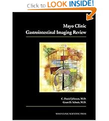
Mayo Clinic Gastrointestinal Imaging Review

Mayo Clinic Gastrointestinal Imaging Review by C. Daniel Johnson & Grant D. Schmit stands as the foremost guide to common diseases and radiographic presentations found within the gastrointestinal tract-authoritatively covering the entire range of gastrointestinal diseases to include conditions affecting the esophagus, stomach, duodenum, small bowel, colon, liver, biliary tree, gallbladder, pancreas, spleen, peritoneum, and mesentery.

Book Details Mayo Clinic Gastrointestinal Imaging Review
  • Paperback: 752 pages
  • Publisher: Informa HealthCare; 1 edition (May 2, 2005)
  • Language: English
  • ISBN-10: 0849397952
  • ISBN-13: 9780849397950
  • Product Dimensions: 10.6 x 8.4 x 1.4 inches
  • List Price: $149.95
  • Price: $132.32
  • You Save: $17.63 (12%)
Buy Now

Copyright 2010 Radiology Book Review - All Rights Reserved.
Designed by Web2feel.com | Bloggerized by Lasantha - Premiumbloggertemplates.com | Affordable HTML Templates from Herotemplates.com.